Bioverfügbares Testosteron beim Mann: Pathophysiologie, die klinische Bedeutung des sexuellen Verlangens, kardiovaskuläre Implikationen und eine Wiederherstellungstherapie mit Testosteron und hCG
Eine klinische Übersichtsarbeit zur evidenzbasierten Präventionspraxis
Zusammenfassung
Die klinische Beurteilung des Androgenstatus des Mannes ist weit über die einfache Messung des Gesamtserums hinausgegangen Testosteron. Gesamtkonzentrationen sind routinemäßig irreführend, da der Großteil des zirkulierenden Testosterons an Plasma gebunden ist Proteine mit völlig unterschiedlichen Affinitäten, wobei das Sexualhormon-bindende Globulin (SHBG) als Hauptregulator dafür dient, wie viel Hormon tatsächlich an das Gewebe abgegeben wird. Diese Übersichtsarbeit fasst das moderne Verständnis von bioverfügbares Testosteron, wobei die Biochemie der Kompartmentierung, die Mechanismen des altersbedingten Rückgangs, die Regulierung von SHBG (einschließlich praktischer Strategien zu dessen Senkung bei klinisch erhöhten Werten), die analytischen Methoden, die zuverlässige Messungen liefern, und die klinische Bedeutung eines verminderten sexuellen Verlangens als spezifischstes klinisches Signal eines echten Androgenmangels behandelt werden. Die kardiovaskulären Implikationen eines niedrigen bioverfügbaren Testosterons werden detailliert untersucht, einschließlich der Ergebnisse der TRAVERSE-Studie und die FDA-Kennzeichnungsaktualisierung von 2025. Der therapeutische Abschnitt befasst sich sowohl mit der Testosteronersatztherapie als auch mit der angemessenen Anwendung von humanem Choriongonadotropin (hCG), das die intratestikuläre Testosteronproduktion und Spermatogenese aufrechtzuerhält. Das Ziel ist ein kohärenter, zeitgemäßer Rahmen, den Kliniker bei männlichen Patienten in der Praxis anwenden können, ohne biochemisches Rauschen mit einer Erkrankung zu verwechseln.
1. Einführung
Ein wachsenes Korpus an Evidenz hat neu definiert, wie Kliniker über Testosteron denken sollten. Das Hormon wirkt nicht proportional zu seiner Gesamtkonzentration im Blut auf das Gewebe; es wirkt proportional zum ungebundenen Anteil plus einem schwach gebundenen Anteil, der sich während der Kapillarpassage von Albumin ablöst. Der Anteil, der fest an SHBG gebunden ist, ist für praktische Zwecke biologisch inert. Dies ist das Wesen der freie Hormon-Hypothese, und obwohl es seit seiner ursprünglichen Formulierung weiterentwickelt wurde, hat sich seine Kernaussage bewährt: Bioverfügbares Testosteron (die Summe aus den freien und den an Albumin gebundenen Fraktionen) ist die aussagekräftigere biologische Variable.1-4
Die klinische Konsequenz ist, dass zwei Männer mit identischen Gesamt-Testosteronkonzentrationen eine sehr unterschiedliche Androgen-Physiologie aufweisen können, wenn sich ihre SHBG-Konzentrationen unterscheiden. SHBG selbst wird durch hepatische Prozesse reguliert, die auf Insulin, Schilddrüsenhormone, Sexualsteroide, entzündliche Zytokine, den Ernährungsstatus und das Altern. Infolgedessen sind häufige Erkrankungen wie Adipositas, Typ 2 Diabetes, Hyperthyreose, Leberfunktionsstörungen und der Alterungsprozess können das Gesamttestosteron von der tatsächlichen Gewebeaktivität des Hormons entkoppeln.5-7
Diese Übersichtsorientiert sich an den Fragen, die ein Kliniker in der Praxis beantworten muss: warum Testosteron mit dem Alter sinkt, warum SHBG ansteigt, worauf es symptomatisch am meisten ankommt (wobei das sexuelle Verlangen der diagnostisch nützlichste Indikator ist), wie das Labor diese Analyten messen sollte, was die kardiovaskuläre Literatur inzwischen sowohl über endogenes Testosteron als auch über die Substitutionstherapie sagt und wie Testosteron und hCG eingesetzt werden sollten, um die Physiologie sicher wiederherzustellen.
2. Kompartimentierung von zirkulierendem Testosteron
2.1 Bindungsfraktionen und ihre biologischen Konsequenzen
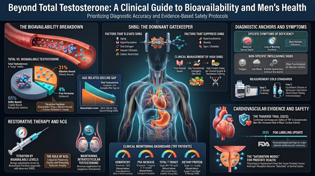
Bei einem gesunden eugonaden Mann sind etwa 44 bis 65 Prozent des Gesamttestosterons an SHBG gebunden, 30 bis 50 Prozent sind lose an Albumin gebunden und 1 bis 4 Prozent zirkulieren als freies Hormon.1,2,3 SHBG ist ein Glykoprotein-Homodimer von etwa 95 kDa, das hauptsächlich von der Leber produziert wird. Seine Assoziationskonstante für Testosteron liegt in der Größenordnung von 1 × 10⁹ pro Molar, was ausreicht, um die an SHBG gebundene Fraktion während der kurzen Passage durch das Kapillarbett effektiv nicht verfügbar zu machen.1 Albumin hingegen ist reichlich vorhanden, bindet Testosteron jedoch mit einer viel geringeren Affinität von etwa 3 × 10⁴ pro Mol.2,3 Die Albuminbindung dissoziiert schnell genug, dass sich der an Albumin gebundene Anteil während der Passage in das Gewebe freisetzen kann, weshalb die Summe aus dem freien und dem an Albumin gebundenen Anteil als bioverfügbares Testosteron bezeichnet wird.2,4
Die Hypothese des freien Hormons besagt, dass nur das ungebundene Molekül durch die Zellmembran diffundiert, um auf den intrazellulären Androgenrezeptor zu wirken.4 Neuere In-vivo-Daten unter Verwendung hochsensitiver Assays haben dieses Bild präzisiert und legen nahe, dass die Dissoziationskinetik von SHBG und Albumin eine gewisse Bioverfügbarkeit über den streng freien Pool hinaus ermöglicht, insbesondere in Geweben mit langsamer kapillärer Passage.2 Die praktische Schlussfolgerung hat sich nicht geändert: Gesamttestosteron allein kann in die Irre führen, und die klinisch aussagekräftige Exposition liegt näher am bioverfügbaren Anteil.
Tabelle 1. Testosteronfraktionen im Plasma bei erwachsenen Männern
| Bruch | Bindungspartner | Affinität (Ka, M⁻¹) | Anteil am Gesamtwert T | Klinische Relevanz |
| Kostenlos | Keine | Nicht anwendbar | 1–4% | Diffundiert frei durch Zellmembranen; aktive Fraktion |
| Albumin-gebunden | Albumin | ~3 × 10⁴ | 30–50% | Locker gebunden; setzt sich während der Kapillarpassage frei und trägt zur Gewebeversorgung bei |
| SHBG-gebunden | SHBG | ~1 × 10⁹ | 44–65% | Fest gebunden; unter normalen Bedingungen biologisch inert |
| bioverfügbar | Frei + an Albumin gebunden | Gemischt | ~30–54% | Bester Index der Gewebeexposition gegenüber Androgenen |
2.2 Warum das Gesamttestosteron nicht ausreicht
Da die an SHBG und an Albumin gebundenen Fraktionen durch Massenwirkungsgleichgewichte bestimmt werden, verschiebt eine Änderung der SHBG-Konzentration die Verteilung zwischen gebundenem und freies Testosteron selbst wenn die Hodenproduktion unverändert bleibt. Eine Verdopplung des SHBG, was beim Übergang vom jungen Erwachsenenalter ins siebte Lebensjahrzehnt nicht ungewöhnlich ist, kann den Gesamt-Testosteronspiegel normalisieren, während der Spiegel des bioverfügbaren Testosterons erheblich gesunken ist.2,9,13 Dies ist der Hauptgrund, warum ältere Männer mit normal erscheinendem Gesamttestosteron klinisch relevante Symptome eines Androgenmangels aufweisen können.
3. Warum der Testosteronspiegel mit dem Alter sinkt
Der altersbedingte Rückgang des Androgenspiegels bei Männern ist mittlerweile gut dokumentiert. Der Baltimore-Längsschnittstudie zum Altern Sie begleiteten gesunde Männer über Jahrzehnte hinweg und berichteten von einem Rückgang des Gesamt-Testosterons um etwa 1 bis 2 Prozent pro Jahr nach dem dritten Lebensjahrzehnt, wobei das freie und das bioverfügbare Testosteron mit etwa 2 bis 3 Prozent pro Jahr stärker abnahmen, da das SHBG parallel dazu ansteigt.9 Die Massachusetts Male Aging Study bestätigte ähnliche Verläufe in einer bevölkerungsbasierten Kohorte.10 Die European Male Ageing Study (EMAS) zeigte zudem, dass bei Männern mit ansonsten normaler Hypophysenfunktion der Rückgang eine Mischung aus testikulären und zentralen Mechanismen widerspiegelt.11,15
3.1 Mechanismen des Rückgangs
Mehrere Signalwege wirken gleichzeitig zusammen, weshalb das Syndrom der spät einsetzender Hypogonadismus ist heterogen.
Hodenalterung. Die Anzahl der Leydig-Zellen nimmt ab, und die überlebenden Leydig-Zellen weisen eine verminderte steroidogene Kapazität auf. Mitochondriale Dysfunktion, Lipofuszin-Akkumulation und eine beeinträchtigte StAR-vermittelte Cholesterin All diese Transportprozesse tragen zu einer geringeren Testosteronproduktion pro Zelle bei.13
Verlust der hypothalamisch-hypophysären Pulsatilität. GnRH-Pulse werden seltener und flacher, LH-Pulse folgen diesem Beispiel, und die zur Stimulation der Testosteronsekretion erforderliche LH-Amplitude wird weniger zuverlässig erreicht. Veldhuis und Kollegen zeigten, dass dieser Verlust der geordneten Pulsatilität selbst eine unabhängige Komponente des reproduktiven Alterns beim Mann ist.14
Veränderung der Körperzusammensetzung. Viszerale Adipositas nimmt im mittleren Lebensalter zu, selbst bei Männern, die ein stabiles Gewicht halten. Fettgewebe schüttet entzündungsfördernde Zytokine aus, die die Sekretion von GnRH und hypophysären Gonadotropinen unterdrücken, und enthält Aromatase, die Testosteron in Östradiol umwandelt. Das Resultat ist ein funktioneller sekundärer Hypogonadismus, der sich dem primären testikulären Altern überlagert.20,21,22
Steigendes SHBG. Die hepatische SHBG-Synthese steigt bei den meisten Männern nach dem dritten Lebensjahrzehnt um etwa ein Prozent pro Jahr an.2,9 Da SHBG Testosteron bindet, führt derselbe Gesamttestosteronspiegel bei älteren Männern zu einem geringeren bioverfügbaren Anteil. Die Travison-Harmonisierungsstudie hat altersstratifizierte Referenzbereiche erstellt, die diese Physiologie berücksichtigen.12
Säkulare Trends. Travison und Kollegen zeigten, dass der Testosteronspiegel auf Bevölkerungsebene bei amerikanischen Männern in den letzten Jahrzehnten über die Geburtskohorten hinweg unabhängig vom Alter gesunken ist, was darauf hindeutet, dass Umweltfaktoren (wahrscheinlich einschließlich der Prävalenz von Adipositas, endokrinen Disruptoren und Veränderungen der körperlichen Aktivität) zusätzlich zum intrinsischen Alterungsprozess eine Rolle spielen.16
3.2 Wie das Altern die Symptome beeinflusst
EMAS stellte fest, dass nur ein begrenztes Spektrum von Symptomen den Androgenstatus bei älteren Männern zuverlässig abbildet. Drei sexuelle Symptome (schlechtere Morgenerektion, geringeres sexuelles Verlangen und erektile Dysfunktion) definieren zusammen mit einem Gesamttestosteron von unter ca. 11 nmol/L (320 ng/dL) und einem freien Testosteron von unter 220 pmol/L (64 pg/mL) den spezifischsten Phänotyp des Hypogonadismus mit späten Beginn.11 Die anderen Symptome, die typischerweise mit einem niedrigen Testosteronspiegel in Verbindung gebracht werden, darunter Müdigkeit, gedrückte Stimmung und verminderte Kraft, sind bei älteren Männern zwar häufig, korrelieren jedoch nur schwach mit dem Androgenstatus.
4. SHBG: Regulation und warum es wichtig ist
SHBG fungiert als der wichtigste „Torwächter“ für Androgene und Östrogen Bioverfügbarkeit. Seine Synthese in der Leber wird durch HNF-4α reguliert, wobei Insulin, Schilddrüsenhormone, Sexualsteroide, Entzündungszytokine und Nährstoffsensoren entweder fördernd oder hemmend auf diesen Prozess einwirken.1,5,6,17 Wichtig ist, dass ein niedriger SHBG-Spiegel mittlerweile als unabhängiger Biomarker von Insulinresistenz sowie bei Typ-2-Diabetes, während ein hoher SHBG-Spiegel ein Indikator für Leber- und Stoffwechselzustände ist, die die Interpretation eines Gesamt-Testosteronwerts beeinflussen.18,19
4.1 Bedingungen, die das SHBG erhöhen
Alterung ist die häufigste Ursache für einen erhöhten SHBG-Spiegel, wobei die oben beschriebene, stetige Aufwärtsverschiebung auftritt. Hyperthyreose erhöht SHBG durch Stimulation von HNF-4α und HNF-1α.17 Östrogenexposition (endogen, durch orale Kontrazeptiva oder durch eine Östrogenersatztherapie) induziert die hepatische SHBG-Synthese deutlich, was ein Mechanismus ist, durch den oral verabreichtes, aber nicht transdermales Östrogen das freie Testosteron bei Frauen senkt.6 Leberzirrhose erhöht häufig den SHBG-Spiegel (manchmal paradoxerweise, in Anbetracht der ansonsten beeinträchtigten hepatischen Synthesefunktion) aufgrund eines veränderten Hepatozytenstoffwechsels und einer verminderten Clearance.24 Anorexia nervosa, schwerer Kalorienmangel und chronischer Proteinmangel erhöhen das SHBG. Mehrere Antikonvulsiva (Phenytoin, Carbamazepin) und einige HIV-Medikamente induzieren SHBG über hepatische Enzymwirkungen.6
4.2 Bedingungen, die SHBG senken
Hyperinsulinämie ist die klinisch bedeutendste Einzelursache für einen niedrigen SHBG-Spiegel in der heutigen Bevölkerung. Selva und Kollegen konnten nachweisen, dass Monosaccharide (insbesondere Fruktose und Glucose) unterdrücken die Transkription von SHBG in der Leber durch eine Herunterregulierung von HNF-4α, wobei die Lipogenese in der Leber als unmittelbarer Vermittler fungiert.5 Das erklärt, warum ein niedriger SHBG-Spiegel so zuverlässig mit Metabolisches Syndrom, Adipositas und Typ-2-Diabetes sowie warum SHBG mittlerweile als Biomarker in der Stoffwechselforschung eingesetzt wird.18,19,40 Weitere Suppressoren umfassen Hypothyreose, exogene Androgene (einschließlich supraphysiologischer Testosteronspiegel, anabole Steroide und Danazol), Glukokortikoidüberschuss, Wachstumshormon- oder IGF-1-Überschuss sowie Gestagene mit androgener Aktivität.6
Tabelle 2. Wichtigste Modulatoren der hepatischen SHBG-Synthese
| Erhöht SHBG | Senkt SHBG |
| Altern (stetiger Anstieg ab dem dritten Lebensjahrzehnt) | Hyperinsulinämie und Insulinresistenz |
| Hyperthyreose | Fettleibigkeit, insbesondere viszerale Adipositas |
| Orale Östrogenexposition (orale Kontrazeptiva, Schwangerschaft) | Typ-2-Diabetes mellitus |
| Leberzirrhose | Hypothyreose |
| Kalorienrestriktion und Anorexia nervosa | Exogene Androgene und anabole Steroide |
| HIV-Infektion (multifaktoriell) | Glucokortikoidüberschuss (Cushing-Syndrom) |
| Antikonvulsiva (Phenytoin, Carbamazepin) | Akromegalie (GH/IGF-1-Überschuss) |
| Chronisch Entzündung (variabler Effekt) | Nephrotisches Syndrom (Proteinverlust über den Urin) |
4.3 Senkung klinisch erhöhter SHBG-Werte durch nicht-pharmakologische Maßnahmen
Wenn SHBG in einem klinisch relevanten Kontext erhöht ist (häufig bei älteren Männern mit relativ schlankem Körperbau, bei subklinischer Hyperthyreose oder bei Männern, die aus beliebigem Grund orale Estrogene einnehmen), besteht die Priorität darin, die zugrundeliegende Ursache zu behandeln. Die folgenden Strategien haben die stärkste mechanistische Unterstützung, obwohl die Evidenzbasis für viele eher beobachtend als randomisiert ist.
Krafttraining sowie eine ausreichende Proteinzufuhr über die Nahrung. Durch körperliche Betätigung hervorgerufene Anstiege bei magere Masse, insbesondere durch Krafttraining, gehen mit einer leichten Senkung des SHBG einher, wahrscheinlich aufgrund einer verbesserten Insulinsensitivität auf Muskelebene. Eine ausreichende Proteinzufuhr (1,2 bis 1,6 g/kg Körpergewicht bei älteren, aktiven Männern) fördert den Aufbau von Muskelmasse und begrenzt den katabolen Zustand, der wiederum zu einem Anstieg des SHBG führt.20,21
Kohlenhydrat Qualität und glykämische Last. Die Wirkung einer Ernährungsumstellung auf SHBG hängt davon ab, welcher Faktor vorherrscht, und muss bei verschiedenen Patienten-Phänotypen separat betrachtet werden. Bei insulinresistenten Männern mit niedrigem SHBG führt eine Verbesserung der Insulinsensitivität (durch Reduzierung von raffiniertem Zucker, mit Fructose gesüßten Getränken und Lebensmitteln mit hoher glykämischer Last) tendenziell dazu, dass SHBG in Richtung Normalwert ansteigt. Bei unterernährten oder energiedefizienten Männern mit hohem SHBG aufgrund von Kalorieneinschränkung führt eine adäquate Kalorienzufuhr tendenziell zu einer Senkung des SHBG. Bei Männern mit hohem SHBG, das auf eine Schilddrüsenerkrankung, die Einnahme von oralem Estrogen, Antikonvulsiva oder eine Lebererkrankung zurückzuführen ist, reicht eine Ernährungsumstellung allein nicht aus; die geeignete Intervention zielt auf die zugrunde thegende Ursache ab.5,18
Behandlung der subklinischen Hyperthyreose. Wenn TSH supprimiert und SHBG hoch ist, normalisiert die Behebung der Schilddrüsenauffälligkeit das SHBG oft innerhalb weniger Monate.17
Reduzierung oder Ersatz von oralem Östrogen. Patientinnen, die aus beliebigen Gründen orale Östrogene einnehmen, können auf transdermale Östrogene umgestellt werden, was den First-Pass-Effekt in der Leber umgeht und nur geringe Veränderungen des SHBG bewirkt.6
Überprüfung der Medikamente. Antikonvulsiva und bestimmte antiretrovirale Medikamente müssen möglicherweise ersetzt werden, wenn ein Hypogonadismus bestätigt ist und SHBG die Hauptursache ist.6
Behandlung von Lebererkrankungen. Bei Männern mit chronischer Lebererkrankung, bei denen das SHBG paradoxerweise erhöht ist, sollte die Behandlung des Leberprozesses (Alkohol Entwöhnung, antivirale Therapie bei Hepatitis C oder Gewichtsverlust (bei Lebersteatose) ist die geeignete Maßnahme.24
Angemessene Kalorienzufuhr. SHBG steigt bei Unterernährung stark an. Sportler und ältere Männer in negativer Energiebilanz können hohe SHBG-Werte aufweisen, die sich durch kalorische Wiederauffüllung normalisieren.6,15
Bor-, Zink- und Magnesium-Supplementierung werden manchmal zur SHBG-Modulation empfohlen. Die klinische Evidenz ist begrenzt und inkonsistent, und keines dieser Mittel ersetzt die Behebung der oben genannten zugrunde liegenden Ursachen.
5. Bedingungen, die das Gesamt-Testosteron vom bioverfügbaren Testosteron entkoppeln
5.1 Adipositas und funktioneller sekundärer Hypogonadismus
Adipositas, insbesondere viszerale Adipositas, ruft ein Muster hervor, das als männliches Adipositas-assoziiertes sekundäres Hypogonadismus-Syndrom (MOSH) bezeichnet wird.22 Dieses Muster ist durch einen niedrigen Gesamt-Testosteronspiegel bei unangemessen normalen oder niedrigen LH- und FSH-Werten gekennzeichnet, was auf eine zentrale Komponente hindeutet. Dabei wirken drei Mechanismen gleichzeitig: Eine Hyperinsulinämie unterdrückt die SHBG-Produktion in der Leber, wodurch der Gesamt-Testosteronspiegel sinkt, ohne dass dabei zwangsläufig der freie Testosteronspiegel sinkt; das viszerale Fettgewebe schüttet IL-6, TNF-α und Leptin in einer Weise, die die GnRH-Ausschüttung im Hypothalamus unterdrückt; außerdem wandelt die Aromatase im Fettgewebe Testosteron in Östradiol um, was eine zusätzliche negative Rückkopplung auf den Hypothalamus bewirkt.20,22,23 Bei Männern mit Diabesity erfüllen in Querschnittsstudien mehr als 30 Prozent die biochemischen Kriterien für Hypogonadismus.23
Entscheidenderweise kehren Gewichtsverlust und eine Verbesserung der Insulinsensitivität dieses Bild oft teilweise oder ganz um. Eine bariatrische Chirurgie kann den Testosteronspiegel bei adipösen, hypogonadnen Männern normalisieren, und selbst ein moderater Gewichtsverlust von 5 bis 10 Prozent verbessert sowohl das Gesamttestosteron als auch das freie Testosteron.20,21,22
5.2 Stoffwechselassoziierte steatotische Lebererkrankung (MASLD)
Fettleber ist eine phänotypische Expression der hepatischen Insulinresistenz und ist eng mit niedrigem SHBG und niedrigem Gesamt-Testosteron assoziiert.19,40 Freies oder bioverfügbares Testosteron ist bei diesen Patienten der aussagekräftigere Messwert. Das Vorliegen einer Steatohepatitis stellt eine zweite Ebene dar, da das Fortschreiten zu Fibrose und Zirrhose paradoxerweise das SHBG erhöhen kann.24
5.3 Leberzirrhose
Fortgeschrittene Zirrhose führt zu einer komplexen Endokrinopathie. SHBG steigt trotz verminderter hepatischer Synthesefunktion in vielen anderen Bereichen an; Albumin fällt ab; die Östradiol-Clearance nimmt ab. Das Gesamt-Testosteron kann trügerisch erhalten erscheinen, während das freie Testosteron eingebrochen ist.24 Die bioverfügbare Messung ist in dieser Population zwingend erforderlich, andernfalls wird die Diagnose verpasst.
5.4 Nephrotisches Syndrom
Eine massive Proteinurie kann zu einem urinären Verlust von SHBG und Albumin führen, wodurch beide Werte sinken. Das Gesamttestosteron sinkt, da die Transportkapazität verringert wurde; das freie Testosteron kann anfangs durch eine Kompensation der HPG-Achse aufrecht erhalten werden, jedoch beeinträchtigen chronischer kataboler Stress und direkte Zytokineffekte auf die Leydig-Zellen schließlich die Produktion.25
5.5 Hyperthyreose und Hypothyreose
Hyperthyreose erhöht SHBG durch Induktion von HNF-1α und HNF-4α, was das Gesamttestosteron erhöht, jedoch geringere Auswirkungen auf das bioverfügbare Testosteron hat.17 Hypothyreose erzeugt das entgegengesetzte Muster. Bei jedem Patienten, bei dem das Gesamttestosteron klinisch interpretiert wird, sollte der Schilddrüsenstatus bekannt sein.
6. Kardiovaskuläre Auswirkungen von bioverfügbarem Testosteron
Die kardiovaskuläre Relevanz des Testosteronstatus ist der Teil dieses Fachgebiets, der sich in den letzten zwei Jahrzehnten am stärksten verändert hat. Ältere Literatur betrachtete Testosteron als kardiovaskulär neutral oder schädlich; die aktuelle Evidenz unterstützt eine nuanciertere Sichtweise, bei der physiologische Spiegel von bioverfügbarem Testosteron mit einem günstigen kardiovaskulären Phänotyp assoziiert sind, während sowohl sehr niedrige als auch supraphysiologische Spiegel Risiken bergen.
6.1 Vaskuläre Biologie von Testosteron
Testosteron hat direkte Wirkungen auf die Blutgefäße. Es fördert die Endothelzelle Stickstoffmonoxid Synthase-Aktivität, die die endothelabhängige Vasodilatation unterstützt. Sie moduliert die Aktivität der Kalziumkanäle in der glatten Gefäßmuskulatur und trägt so zur koronaren und peripheren Vasodilatation bei. Sie beeinflusst den Entzündungsstatus in der Gefäßwand, wobei in vielen Kohorten ein niedriger Testosteronspiegel mit erhöhten CRP- und IL-6-Werten in Verbindung gebracht wird.34,50,56 Auf der Ebene der Kardiomyozyten werden Androgenrezeptoren exprimiert, und eine physiologische Androgenversorgung scheint für einen normalen Energiestoffwechsel des Myokards permissiv zu sein.
Testosteron steht zudem in Wechselwirkung mit dem Lipoprotein System in einer Weise, die für das ASCVD-Risiko klinisch relevant ist. Supraphysiologische Testosteronspiegel, einschließlich der meisten Formen des Missbrauchs an anabolen Steroiden, senken HDL und kann zu einem Anstieg der ApoB-haltigen Lipoproteine führen. Eine physiologische Substitution führt dagegen in der Regel zu einer leichten Senkung der Gesamtcholesterin und ApoB sowie eine leichte Senkung des HDL-Spiegels, mit neutralen oder positiven Auswirkungen auf die Insulinsensitivität und viszerales Fett.49,50,56 Die Auswirkungen auf Plaque Die Biologie, die sich aus der Wiederherstellung der normalen Physiologie ergibt – im Gegensatz zu einer überphysiologischen Exposition –, ist die Variable, die für die Prävention entscheidend ist.
6.2 Endogenes Testosteron und kardiovaskuläre Endpunkte
Groß prospektive Kohorten Es wurde allgemein festgestellt, dass ein niedriger endogener Testosteronspiegel mit einem erhöhten Risiko für kardiovaskuläre Erkrankungen und Gesamtmortalität bei Männern. Die gepoolte Analyse von Yeap und Kollegen, in der 11 prospektive Kohorten mit insgesamt über 24.000 Männern zusammengefasst wurden, ergab, dass Männer im untersten Quintil des Gesamt-Testosteronspiegels ein höheres Risiko für kardiovaskuläre Ereignisse aufwiesen und dass ein sehr niedriger freier Testosteronspiegel (unter etwa 184 pmol/l, bzw. 53 pg/ml) das größte Risiko darstellte.34 A Meta-Analyse Corona und Kollegen stellten ähnliche inverse Zusammenhänge zwischen endogenem Testosteron und kardiovaskulären Ereignissen fest.35 Mendelsche Randomisierung Analysen haben zwar eher gemischte Ergebnisse geliefert, stützen jedoch im Allgemeinen nicht die Annahme, dass physiologisches Testosteron einen schädlichen kausalen Einfluss auf kardiovaskuläre Endpunkte hat, und einige Analysen deuten auf einen schützenden Zusammenhang am unteren Ende der Verteilung hin.55
SHBG selbst scheint ein unabhängiger kardiovaskulärer und metabolischer Biomarker zu sein. Ein niedriges SHBG ist ein Prädiktor für Typ-2-Diabetes,18 und ein sehr hoher SHBG-Spiegel bei älteren Männern wurde in einigen Kohorten mit der Mortalität in Verbindung gebracht, obwohl die Interpretation durch umgekehrte Kausalität (Gebrechlichkeit und Unterernährung führen zu einem Anstieg des SHBG).19,58
6.3 Testosteron-Substitution und kardiovaskuläres Risiko: Aktuelle Evidenz nach TRAVERSE
In den Jahren 2013 und 2014 berichteten zwei viel beachtete Studien (Vigen et al. im VA-System und Finkle et al. in einer Abrechnungsdatenbank) über Zusammenhänge zwischen Testosteron-Ersatztherapie (TRT) und kardiovaskulären Ereignissen. Beide wiesen schwerwiegende methodische Einschränkungen auf, darunter die unvollständige Erfassung der Testosteronüberwachung, kurze Nachbeobachtungszeiten und Selektionsprobleme. Nachfolgende Analysen warfen methodische Bedenken hinsichtlich beider Studien auf.54, 54b Trotzdem fügte die FDA im Jahr 2015 einen Warnhinweis in die Produktinformation ein, der vor möglichen kardiovaskulären Risiken warnte, was zu einem Rückgang der Verschreibungen führte.
Seitdem haben mehrere Beobachtungsstudien an genauer definierten Populationen neutrale oder positive Zusammenhänge festgestellt. Sharma und Kollegen stellten in einer VA-Kohorte mit mehr als 83.000 Männern fest, dass die Normalisierung des Testosteronspiegels durch eine Testosteronersatztherapie (TRT) mit einer verringerten Inzidenz von Myokardinfarkt, Schlaganfall, sowie die Gesamtmortalität im Vergleich zu Männern, deren Werte niedrig blieben oder die nicht behandelt wurden.37 Cheetham und Kollegen berichteten in Daten von Kaiser Permanente über ein um 33 Prozent geringeres Risiko für kardiovaskuläre Ereignisse bei TRT-behandelten Männern.38 Anderson und Kollegen berichteten im Intermountain-Healthcare-System ebenfalls über neutrale oder günstige kardiovaskuläre Ergebnisse.39
Die bislang größte randomisierte Studie zur kardiovaskulären Sicherheit war TRAVERSE (Lincoff et al., New England Journal of Medicine, 2023), eine randomisierte, placebokontrollierte Nichtunterlegenheitsstudie mit etwa 5.200 Männern im Alter von 45 bis 80 Jahren mit Hypogonadismus und entweder bereits bestehender Herz-Kreislauf-Erkrankung oder einem hohen kardiovaskulären Risiko. Während einer mittleren Nachbeobachtungszeit von 22 Monaten trat das primäre Endpunktkombinat aus kardiovaskulärem Tod, nicht tödlichem Myokardinfarkt und nicht tödlichem Schlaganfall bei 7,0 Prozent der Testosteron-Gruppe und bei 7,3 Prozent der Placebo Gruppe, die das vorab festgelegte Kriterium für die Nichtunterlegenheit erfüllt (Hazard-Ratio 0,96; 95%-Konfidenzintervall 0,78 bis 1,17).36 Vorhofflimmern, akutes Nierenversagen und Lungenembolie traten in der Testosteron-Gruppe etwas häufiger auf, was mit den bekannten biologischen Mechanismen der durch Testosteron verursachten Erythrozytose; dies sind die wichtigsten verbleibenden Sicherheitsparameter, die überwacht werden müssen. TRAVERSE war eine Nichtunterlegenheitsstudie mit moderater Nachbeobachtungsdauer, die keinen langfristigen kardiovaskulären Nutzen nachweist und auch nicht umfassend auf die Plaque-Biologie eingeht, Blutdruck, oder eine breitere Anwendung außerhalb der zugelassenen Indikationen.
Im Jahr 2025 ordnete die FDA klassenweite Kennzeichnungsänderungen für Testosteronprodukte an. Die aktualisierten Etiketten enthielten die Ergebnisse der TRAVERSE-Studie und entfernten den Warnhinweis (Boxed Warning) bezüglich eines erhöhten Risikos schwerwiegender unerwünschter kardiovaskulärer Ereignisse. Die überarbeiteten Etiketten fügten Warnhinweise hinsichtlich Blutdruckerhöhung hinzu oder verschärften diese und behielten die bestehenden Anwendungseinschränkungen für altersbedingten Hypogonadismus bei fehlendem Nachweis eines biochemischen Mangels bei. Ärzte sollten die aktuellen Verschreibungsinformationen für das jeweils verwendete Präparat heranziehen.
6.4 Testosteron und Koronarplaque
Die kardiovaskuläre Teilstudie der „Testosterone Trials“ (Budoff et al., JAMA, 2017) berichtete, dass bei Männern, die ein Jahr lang Testosteron-Gel erhielten, ein stärkerer Anstieg der nicht verkalkten Koronararterien zu verzeichnen war Plaquevolumen auf Koronare CT-Angiographie als die Placebo-Empfänger.48 Die Studie war hinsichtlich klinischer Ereignisse nicht auf eine ausreichende Aussagekraft ausgelegt; der Anstieg bei nicht verkalkte Plaque war bescheiden, und die längerfristigen klinischen Auswirkungen bleiben ungewiss. Im Zusammenhang mit der TRAVERSE-Studie ist die plausibelste Interpretation, dass die physiologische Wiederherstellung bei moderater Nachbeobachtungsdauer nachweislich nicht zu einer Zunahme klinischer kardiovaskulärer Ereignisse geführt hat, während die Endpunkte der Plaque-Bildgebung Anzeichen liefern, die eine fortgesetzte Überwachung rechtfertigen.
6.5 Erythrozytose und thrombotisches Risiko
Das am konsistentesten reproduzierbare kardiovaskuläre Nebenwirkungssignal der TRT ist die Erythrozytose. Testosteron stimuliert die Erythropoese sowohl durch direkte Wirkungen auf das Knochenmark als auch durch eine erhöhte Erythropoietinproduktion, wobei das Ausmaß der Wirkung von der Darreichungsform abhängt. Injizierbare Ester bewirken bei Erreichen des Spitzenwerts den stärksten Anstieg; transdermale Präparate den geringsten. A Hämatokrit Ein Wert über 54 Prozent gilt allgemein als Hinweis darauf, die Dosis zu reduzieren, die Darreichungsform zu ändern oder die Therapie vorübergehend auszusetzen und zu prüfen, ob Störvariablen wie beispielsweise obstruktiv Schlafapnoe und Dehydrierung.7,54
6.6 Synthese für den Herz-Kreislauf-Praktiker
Ein endogenes Testosteronspiegel am unteren Ende des physiologischen Bereichs ist mit einem erhöhten kardiovaskulären Risiko verbunden, was mit ziemlicher Sicherheit auf eine Kombination aus echter biologischer Anfälligkeit und verwirrend durch einen ungünstigen metabolischen Phänotyp. Es wurde nicht nachgewiesen, dass die Wiederherstellung eines Testosteronspiegels im physiologischen Bereich bei symptomatischen Männern mit Hypogonadismus – bei angemessener Überwachung – zu einer Erhöhung führt schwerwiegende unerwünschte kardiovaskuläre Ereignisse während der untersuchten Nachbeobachtungszeiträume. Eine Erythrozytose muss überwacht werden. Intensive Behandlung herkömmlicher kardiovaskulärer Risikofaktoren (Senkung des ApoB-Spiegels, Blutdruckkontrolle, Blutzuckereinstellung, Rauchen (Raucherentwöhnung und körperliche Aktivität) bleibt die wichtigste kardiovaskuläre Maßnahme. Die Testosteronersatztherapie ist eine ergänzende Maßnahme, die einen bestimmten Mangel behebt, und stellt keine eigenständige kardiovaskuläre Therapie dar.
7. Sexuelles Verlangen: Das diagnostisch nützlichste Symptom eines Testosteronmangels
Von allen Symptomen, die einem niedrigen Testosteronspiegel zugeschrieben werden, ist das nachlassende sexuelle Verlangen dasjenige, das am zuverlässigsten und spezifischsten mit dem Androgenstatus zusammenhängt. Dies ist kein unwesentliches Detail; es ist der nützlichste diagnostische Anker, den ein Kliniker bei der Sichtung der unspezifischen Beschwerden hat, die oft mit Alterung und Stoffwechselerkrankungen einhergehen. Es ist wichtig anzumerken, dass die Endocrine Society empfiehlt, einen Hypogonadismus nur bei Männern zu diagnostizieren, die Symptome aufweisen, die mit einem Testosteronmangel übereinstimmen, und deren Testosteronkonzentrationen eindeutig und konsistent niedrig sind, was durch mindestens zwei getrennte Morgenmessungen bestätigt wurde; der Symptomstatus allein reicht nicht aus.7
7.1 Warum ein vermindertes sexuelles Verlangen das diagnostisch nützlichste Symptom ist
Travison und Kollegen zeigten in einer Analyse der Massachusetts Male Aging Study einen abgestuften Zusammenhang zwischen bioverfügbarem Testosterone und der Libido, der über den gesamten physiologischen Bereich hinweg besteht, während die Beziehung zwischen Testosteron und der erektilen Funktion deutlich weniger konsistent ist.29 EMAS identifizierte unabhängig drei sexuelle Symptome (mangelnde Morgenerektion, geringe sexuelle Lust und erektile Dysfunktion) als die einzigen Symptome, die niedriges Testosteron mit vertretbarer Spezifität anzeigten, sobald das Alter berücksichtigt wurde.11 Von den dreien ist geringes Verlangen am spezifischsten für den Androgenstatus; erektile Dysfunktion wird stark durch vaskuläre, neurogene und pharmakologische Ursachen beeinträchtigt, und Morgen Erektionen sind ein sensitiver, aber weniger spezifischer Indikator.
In randomisierten Testosteron-Ersatztherapie-Studien ist die konsistenteste und größte Effektstärke beim sexuellen Verlangen zu beobachten. Die Sexualfunktion-Teilstudie der Testosteron-Studie (Cunningham et al., 2016) zeigte, dass von den drei primären sexuellen Zielparametern der Anstieg der Werte für das sexuelle Verlangen sowohl bei den Männern am konsistentesten als auch klinisch am bedeutsamsten war.30 Meta-analysen zu den Effekten einer Testosteronersatztherapie auf die Sexualfunktion zeigen ebenfalls, dass die Libido das vorherrschende Signal ist, mit geringeren und variableren Effekten auf die erektile Funktion.32
7.2 Was Patienten beschreiben und was zu fragen ist
Männer mit androgenbedingtem vermindertem Libido beschreiben einen grundlegenden Rückgang des spontanen Interesses, das zuvor sexuelle Gedanken und Verhaltensweisen auslöste. Sie stellen fest, dass sie nicht mehr unaufgefordert über Sex nachdenken, gewöhnliche Reize nicht mehr als erotisch bedeutsam empfinden, und beschreiben die Veränderung oft eher als Verlust eines inneren Antriebs denn als Verlust der Fähigkeit. Männer können in der Regel angeben, ob sich ihre Libido im Vergleich zu ihrem eigenen früheren Ausgangswert verändert hat, was diagnostisch aussagekräftiger ist als bevölkerungsbezogene Schwellenwerte.
Der Verlust nächtlicher und frühmorgendlicher Erektionen ist eng miteinander verbunden und wird häufig gemeinsam berichtet. Das klinische Gespräch sollte sowohl explizit nach der Libido als auch nach morgendlichen Erektionen fragen. Die alleinige Frage nach der Erektionsfähigkeit ruft Antworten hervor, die vaskuläre, neurologische, pharmakologische und androgene Beiträge vermischen; die gezielte Frage nach Verlangen und morgendlichen Erektionen trennt diese voneinander.
7.3 Was sexuelle Symptome nicht vorhersagen
Eine erektile Dysfunktion ohne verminderten Sexualtrieb lässt sich selten allein durch eine Testosteronersatztherapie beheben. Bei Männern mit isolierter erektiler Dysfunktion und eindeutig normalem bioverfügbarem Testosteron stehen Gefäßerkrankungen im Vordergrund der Differentialdiagnose (oftmals das erste Anzeichen einer generalisierten Atherosklerose), neurogene Ursachen, antihypertensiv Medikamente, Antidepressiva und psychologische Faktoren. Eine Behandlung dieser Patienten mit Testosteron ist selten wirksam und setzt sie den Nebenwirkungen einer Hormonersatztherapie aus, ohne dass ein entsprechender Nutzen entsteht.32,33
8. Weitere Manifestationen des Androgenmangels
Über sexuelle Symptome hinaus umfasst das Testosteronmangelsyndrom körperliche, metabolische und psychologische Merkmale. Keines davon ist so spezifisch wie eine verminderte Libido, aber im richtigen klinischen Kontext untermauern sie die Diagnose.
8.1 Körperzusammensetzung und Stoffwechsel
Männer mit Hypogonadismus bilden viszerales Fettgewebe an und verlieren fettfreie Muskelmasse, was mit einem entsprechenden Rückgang der Kraft einhergeht.20,21,49,60 Dies ist zum einen eine direkte Folge des Verlusts der Androgen-Signalübertragung in Muskel- und Fettgewebe und zum anderen eine Folge des metabolischen Syndroms, in dessen Rahmen sich ein durch Adipositas bedingter Hypogonadismus entwickelt.59 Langzeitstudien zur Hormonersatztherapie bei adipösen Männern mit Hypogonadismus haben eine signifikante Verringerung von Taillenumfang, Körpergewicht und HbA1c; diese Effekte sind oft geringer als die, die mit einer intensiven Lebensstilintervention erzielt werden, scheinen sich jedoch zu diesen hinzuzufügen.49
8.2 Knochen
Testosteron trägt – unter anderem durch die Aromatisierung zu Östradiol – zur Erhaltung der Knochenmineraldichte bei Männern bei. Die Knochen-Teilstudie der Testosteron-Studie zeigte bei älteren Männern mit niedrigem Testosteronspiegel nach einem Jahr Substitutionstherapie signifikante Zuwächse bei der volumetrischen Knochendichte und der geschätzten Knochenfestigkeit.46
8.3 Anämie
Eine leichte Anämie tritt bei unbehandeltem Hypogonadismus häufig auf. Die Teilstudie zur Anämie im Rahmen der „Testosterone Trial“-Studie zeigte, dass sich die Anämie bei einem nennenswerten Anteil der behandelten Männer zurückbildete, darunter auch bei einigen, bei denen keine andere Ursache festgestellt werden konnte.47
8.4 Stimmung, Motivation und Kognition
Viele Männer mit Hypogonadismus berichten über Niedergeschlagenheit, Reizbarkeit und einen verminderten Energiepegel; diese Symptome bessern sich in der Regel leicht durch eine Hormonersatztherapie, obwohl sie auch viele andere Ursachen haben können und nicht allein als Grundlage für eine Diagnose herangezogen werden sollten.7,33 In randomisierten Studien waren die kognitiven Effekte einer Hormonersatztherapie bei älteren Männern mit leichten Gedächtnisbeschwerden gering und uneinheitlich.57
9. Messung: Methoden, die funktionieren, und Methoden, die in die Irre führen
9.1 Gesamt-Testosteron
Die Endocrine Society und das Hormonstandardisierungsprogramm der CDC empfehlen seit über einem Jahrzehnt LC-MS/MS als Referenzmethode für die Bestimmung des Gesamt-Testosterons, insbesondere bei den für die Diagnose relevanten niedrigen Konzentrationen. Ältere Plattform-Immunoassays sind im mittleren bis hohen Bereich bei erwachsenen Männern zuverlässig, weisen jedoch bei niedrigen Konzentrationen erhebliche Fehler auf und werden bei Frauen nicht empfohlen.26,27
Präanalytische Variablen spielen eine wichtige Rolle. Der Gesamt-Testosteronspiegel weist einen ausgeprägten morgendlichen Höchstwert und einen reduzierten nachmittäglichen Tiefstwert auf, wobei die tageszeitliche Schwankungsbreite bei älteren Männern abgeschwächt ist. Die Probenentnahme sollte zwischen 7 und 10 Uhr morgens in nüchternem Zustand und an mindestens zwei verschiedenen Tagen erfolgen, bevor die Diagnose eines Hypogonadismus bestätigt wird. Akute Erkrankungen, kürzlich durchgeführte anstrengende körperliche Betätigung und eine kürzlich erfolgte Kalorienrestriktion können den Testosteronspiegel vorübergehend senken und sollten vor der Interpretation eines isolierten niedrigen Wertes berücksichtigt werden.
9.2 Freies Testosteron: Methoden, nach Rangfolge
Direkte analoge Immunoassays zur Bestimmung von freiem Testosteron sind unzuverlässig und werden von den führenden Gesellschaften für Endokrinologie und Andrologie ausdrücklich nicht empfohlen.26,27 Sie können Werte liefern, die um ein Vielfaches vom tatsächlichen freien Anteil abweichen, wobei die Fehler in den Populationen am größten sind, in denen die Messung am wichtigsten ist (Männer mit abnormalen SHBG-Werten).
Die Gleichgewichtsdialyse mit anschließender LC-MS/MS ist die Referenzmethode. Die Serumprobe wird über eine semipermeable Membran gegen einen Puffer dialysiert; dabei gelangt nur das ungebundene Hormon hindurch; das im Dialysat enthaltene Testosteron wird anschließend mittels Massenspektrometrie gemessen. Dies ist der Goldstandard, jedoch arbeitsintensiv und nicht flächendeckend verfügbar.3,28
Das anhand der Vermeulen-Formel berechnete freie Testosteron weist eine hervorragende Übereinstimmung mit der Gleichgewichtsdialyse auf (Korrelationskoeffizient r ≈ 0,99 in sorgfältig durchgeführten Vergleichen), vorausgesetzt, dass das Gesamttestosteron und das SHBG mit präzisen Messsystemen bestimmt werden.3,28 Diese Berechnung ist die praktische Methode, die die meisten Ärzte anwenden sollten; das neuere mehrstufige allosterische Modell von Goldman et al. verbessert die Genauigkeit bei extremen SHBG-Werten, ist jedoch noch nicht routinemäßig verfügbar.2
Tabelle 3. Methoden zur Messung oder Schätzung des freien Testosterons
| Methode | Genauigkeit | Praktische Anwendung | Einschränkung |
| Direkter analoger Immunoassay | Schlecht | Nicht empfohlen | Bei niedrigen und hohen SHBG-Werten erheblich ungenau; die größten Fehler treten in den Bevölkerungsgruppen auf, in denen die Messung am wichtigsten ist |
| Gleichgewichtsdialyse + LC-MS/MS | Referenzstandard | Forschung und diagnostisch schwierige Fälle | Arbeitsintensiv, zeitaufwendig, teuer und in routinemäßigen klinischen Labors nicht weit verbreitet |
| Vermeulen berechnete das freie T | Hoch (r ≈ 0,99 im Vergleich zur Gleichgewichtsdialyse) | Routinemäßiger klinischer Einsatz | Hängt von der Qualität der Gesamt-Testosteron- und SHBG-Werte ab; bei extremen SHBG-Werten weniger genau |
| Goldman-Mehrstufen-Allosterikmodell | Am höchsten bei extremen SHBG-Werten | Ausgewählte schwierige Fälle | In routinemäßigen klinischen Laborsystemen noch nicht flächendeckend eingeführt |
10. Wiederherstellung des bioverfügbaren Testosterons: Pharmakologie
10.1 Therapeutische Ziele
Das Ziel der Hormonersatztherapie besteht darin, die Symptome (mit dem sexuellen Verlangen als primärem Endpunkt) wiederherzustellen und gleichzeitig die biochemischen Werte im physiologischen Bereich gesunder junger Männer zu halten. Die Leitlinie der Endocrine Society sieht einen Gesamt-Testosteronspiegel im mittleren Normalbereich vor, typischerweise 400 bis 700 ng/dL (14 bis 24 nmol/L), mit einem entsprechenden freien Testosteronspiegel im mittleren Normalbereich.7 Bei Männern mit erhöhten SHBG-Werten müssen die Zielwerte für das Gesamt-Testosteron mit Vorsicht interpretiert werden; die Dosisanpassung sollte bei dieser Gruppe auf der Grundlage des bioverfügbaren Testosterons und nicht des Gesamt-Testosterons erfolgen.
10.2 Formulierungen
Topische Gele und Lösungen sorgen für konstante Serumspiegel, die dem physiologischen Tagesrhythmus nahekommen. Aus diesem Grund gelten sie in vielen Leitlinien als Mittel der ersten Wahl, wobei die Nachteile in der täglichen Anwendung, leichten Hautreaktionen und dem geringen, aber realen Risiko einer Übertragung von Mensch zu Mensch liegen.7
Intramuskuläre Ester (Testosteroncypionat oder -enanthat) sind kostengünstig und wirksam. Die klassische Dosierung von 100 bis 200 mg alle 1 bis 2 Wochen führt zu starken Schwankungen; viele Ärzte bevorzugen mittlerweile kleinere, häufigere Injektionen (beispielsweise 50 bis 80 mg zweimal wöchentlich, oft subkutan), um das pharmakokinetische Profil abzuflachen und Schwankungen des Hämatokritwerts zu verringern.7,8
Langwirksames Testosteronundecanoat, das nach einer Initialphase alle 10 bis 14 Wochen durch eine tiefe intramuskuläre Injektion verabreicht wird, sorgt für stabile Spiegel und wird außerhalb der Vereinigten Staaten weit verbreitet eingesetzt. Die US-amerikanische Darreichungsform erfordert aufgrund des historisch seltenen Risikos einer pulmonalen Ölmikroembolie eine Beobachtung in der Arztpraxis.51
Subkutane Testosteron-Pellets, die alle 3 bis 6 Monate implantiert werden, sorgen für einen stabilen Hormonspiegel bei Männern, die eine seltenere Dosierung bevorzugen.8
Oral verabreichtes Testosteronundecanoat (das in den derzeit in den USA erhältlichen Formulierungen über den lymphatischen Weg resorbiert wird) und intranasales Testosteron-Gel sind weitere Optionen, für die jeweils spezifische Überwachungsaspekte zu beachten sind.
10.3 Dosierung bei hohem bzw. niedrigem SHBG-Spiegel
Bei Männern mit hohem SHBG-Spiegel (häufig ältere, schlanke Männer, Männer mit subklinischer Hyperthyreose oder solche, die Antikonvulsiva einnehmen) können höhere Dosen erforderlich sein, um einen zufriedenstellenden Spiegel an bioverfügbarem Testosteron zu erreichen, und der Gesamt-Testosteronspiegel muss möglicherweise im oberen Normbereich liegen. Bei Männern mit niedrigem SHBG-Spiegel (typischerweise adipös oder insulinresistent) können Standarddosen zu einem überhöhten Gehalt an freiem Testosteron führen, wenn die Dosierung ausschließlich anhand des Gesamt-Testosteronspiegels erfolgt, was zu Akne, Reizbarkeit und einer beschleunigten Erythrozytose führen kann. In beiden Patientengruppen sollte die Dosierung anhand des berechneten bioverfügbaren Testosterons erfolgen.
11. Humanes Choriongonadotropin: Wirkmechanismus und klinische Anwendung
Humanes Choriongonadotropin (hCG) wirkt als Analogon des luteinisierenden Hormons (LH), bindet an den LH-Rezeptor auf den Leydig-Zellen und stimuliert direkt die intratestikuläre Testosteronproduktion. Da exogenes Testosteron die Ausschüttung von LH und FSH durch die Hypophyse unterdrückt und die endogene Stimulation der Leydig-Zellen unterbindet (was zur Folge hat, dass der Testosteronspiegel im Hoden sinkt und die Spermatogenese gehemmt wird), spielt hCG zwei wichtige klinische Rollen: als Monotherapie bei ausgewählten Männern und als Ergänzung zur Testosteronersatztherapie, wenn die Erhaltung der Fruchtbarkeit im Vordergrund steht.
11.1 Das Problem des intratestikulären Testosterons
Die Spermatogenese erfordert intratestikuläre Testosteronkonzentrationen, die etwa 50- bis 100-mal höher sind als der Testosteronspiegel im Blutkreislauf. Exogenes Testosteron unterdrückt den LH-Spiegel und beseitigt damit den Reiz für die intratestikuläre Testosteronproduktion. Coviello und Kollegen zeigten, dass niedrig dosiertes hCG (250 IE subkutan jeden zweiten Tag), das gleichzeitig mit exogenem Testosteron verabreicht wird, den Testosteronspiegel im Hoden im Normbereich hält, während Testosteron allein diesen um mehr als 90 Prozent senkt.41
Hsieh und Kollegen zeigten anschließend, dass bei Männern, die eine Testosteronersatztherapie mit gleichzeitiger Gabe von niedrig dosiertem hCG (in der Regel dreimal wöchentlich 500 IE) erhielten, die Spermienparameter während der gesamten Behandlung stabil blieben, während bei denjenigen ohne hCG die erwartete Oligospermie oder Azoospermie auftrat.42 Ramasamy und andere haben die praktische Umsetzung der fruchtbarkeitserhaltenden Kombinationstherapie zusammengefasst.43
11.2 hCG-Monotherapie
Bei jüngeren Männern mit sekundärem oder gemischtem Hypogonadismus, die ihre Fruchtbarkeit erhalten möchten, ist eine hCG-Monotherapie eine sinnvolle erste Wahl. Die typische Dosierung beginnt bei 1.000 bis 1.500 IE subkutan zwei- bis dreimal pro Woche und wird entsprechend der Reaktion von Gesamt-Testosteron, freiem Testosteron und Östradiol titriert. Viele Männer erreichen unter der Monotherapie einen im mittleren Normbereich liegenden Gesamttestosteronspiegel. Der Östradiolspiegel kann stärker ansteigen als bei einer Testosteronersatztherapie (da die Aromatisierung im Hoden erhalten bleibt), und gelegentlich ist der gezielte Einsatz eines Aromatasehemmers gerechtfertigt; dies sollte jedoch nicht reflexartig erfolgen.41,43,44
Die Anwendung von hCG in diesem Zusammenhang erfolgt in den meisten Ländern in der Regel außerhalb der zugelassenen Indikation und sollte individuell angepasst werden, idealerweise unter Einbeziehung der Reproduktionsurologie, wenn die Fertilität ein aktives Ziel ist. Die Evidenz für hCG-Protokolle stammt in erster Linie aus mechanistischen Studien und kleinen Beobachtungs- oder prospektiven Studien und weniger aus großen randomisierte kontrollierte Studien.
11.3 hCG als Ergänzung zur Testosteronersatztherapie
Bei Männern, die eine Testosteronersatztherapie erhalten und ihre Fruchtbarkeit erhalten möchten oder unter einer störenden Hodenatrophie leiden, wird dem Testosteron-Behandlungsschema eine niedrig dosierte hCG-Gabe (in der Regel 500 IE zwei- bis dreimal wöchentlich) hinzugefügt. Diese Dosis erhält den Testosteronspiegel im Hoden und die Spermatogenese aufrecht, ohne zu einem übermäßigen Anstieg des Gesamt-Testosterons oder des Östradiols zu führen.41,42,43 Wie bei der Monotherapie erfolgt diese Kombinationsanwendung in der Regel außerhalb der zugelassenen Indikation, und die zugrunde liegenden Erkenntnisse stammen größtenteils aus Beobachtungsstudien.
11.4 Protokolle zur Wiederaufnahme nach Absetzen von Testosteron
Männer, die exogenes Testosteron eingenommen haben (sei es verschreibungspflichtig oder im Rahmen der Einnahme von anabolen Androgenen) und die ihre endogene Funktion sowie ihre Fruchtbarkeit wiederherstellen möchten, kommen für Wiederherstellungsprotokolle in Frage. Zu den Bestandteilen gehören in der Regel hCG zur Förderung der Regeneration der Leydig-Zellen, ein selektiver Östrogenrezeptormodulator (Clomifen oder Tamoxifen) zur Aufhebung der Hemmung der Gonadotropinsekretion durch die Hypophyse sowie in einigen Fällen rekombinantes FSH, falls sich die Spermatogenese nicht erholt. Wenker und Kollegen berichteten über vielversprechende Erholungsraten bei solchen Kombinationen bei Männern, die nach einer Exposition gegenüber exogenen Androgenen versuchen, ihre Fruchtbarkeit wiederherzustellen.44,45
11.5 Praktische Überlegungen
hCG wird durch subkutane Injektion verabreicht. Die Nebenwirkungen sind in der Regel mild und umfassen lokale Reaktionen an der Injektionsstelle, eine leichte Gynäkomastie (bei steigendem Östradiolspiegel) sowie Akne. Die Kosten waren in der Vergangenheit schwankend, und in einigen Ländern sind individuell zusammengestellte Präparate erhältlich, wobei die Qualitätskontrolle ein berechtigtes klinisches Anliegen darstellt.
12. Überwachung der Therapie
Die Einleitung der Therapie und die Titration erfordern eine systematische Überwachung. Der folgende Rahmen entspricht den Leitlinien der Endocrine Society und der AUA, angepasst an die oben dargelegten kardiovaskulären Erkenntnisse.7,8
Ausgangswert. Gesamttestosteron (LC-MS/MS oder validierter Plattform-Assay), SHBG, berechnetes freies oder bioverfügbares Testosteron (Vermeulen), Albumin, falls die Berechnung durchgeführt werden soll, LH und FSH (zur Unterscheidung zwischen primärem und sekundärem Hypogonadismus), Prolaktin (in ausgewählten Fällen), TSH, großes Blutbild (unter Berücksichtigung des Hämatokrits), umfassendes Stoffwechselprofil, Nüchtern-Lipidprofil einschließlich ApoB, sofern verfügbar, PSA bei Männern über 40 sowie digitale rektale Untersuchung gemäß den AUA-Leitlinien.
Drei Monate. Überprüfung der Symptome (mit dem sexuellen Verlangen als primärem Endpunkt), erneute Bestimmung des Gesamt-Testosterons (zeitlich korrekt auf die Darreichungsform abgestimmt: bei intramuskulären Estern zum Zeitpunkt des Tiefstwerts, bei Gelen 2 bis 8 Stunden nach der Anwendung), großes Blutbild (CBC) und gegebenenfalls PSA. Ein Hämatokritwert über 54 Prozent führt zu einer Dosisreduktion oder einem Wechsel der Darreichungsform.
Sechs und zwölf Monate. Dieselbe Untersuchung, ergänzt um eine erneute Bestimmung der Körperzusammensetzung und des Taillenumfangs sowie der Lipidwerte und der glykämischen Indizes bei Männern mit Stoffwechselerkrankungen.
Danach jährlich. Symptomabklärung, Gesamt- und freies Testosteron, Hämatokrit, PSA bei Männern über 40 sowie eine umfassende Neubewertung des kardiovaskulären Risikos. Ein Anstieg des PSA-Werts um mehr als 1,4 ng/ml innerhalb von 12 Monaten oder ein bestätigter PSA-Wert über 4,0 ng/ml rechtfertigen eine urologische Abklärung.7,8,53
13. Sicherheit: Was zu beachten ist und was bereits geklärt wurde
13.1 Prostata
Aktuelle Daten stützen nicht die ältere Annahme, dass eine physiologische Testosteronersatztherapie bei Männern mit normaler Prostatagesundheit im Ausgangszustand Prostatakrebs verursacht. Das von Morgentaler und Kollegen vorgeschlagene Sättigungsmodell, bei dem die Androgenrezeptor-Signalübertragung im physiologischen Bereich gesättigt ist, passt besser zu den verfügbaren Daten als das lineare Stimulationsmodell, das den älteren Empfehlungen zugrunde liegt.52,53 Das bedeutet nicht, dass eine Hormonersatztherapie bei unbehandeltem Prostatakrebs unbedenklich ist; es bedeutet vielmehr, dass eine unkomplizierte benigne Prostatahyperplasie und ein stabiler PSA-Wert keine Kontraindikation für eine Hormonersatztherapie darstellen, wenn ein symptomatischer Hypogonadismus nachgewiesen ist.
13.2 Erythrozytose
Siehe Abschnitt 6.5. Die Erythrozytose ist die häufigste dosislimitierende Laborabweichung bei der TRT. Bei Männern mit anhaltender Erythrozytose trotz angemessener Dosierung sollte auf eine gleichzeitig bestehende obstruktive Schlafapnoe untersucht werden.
13.3 Fruchtbarkeit
Exogenes Testosteron unterdrückt zuverlässig die Spermatogenese. Männer im gebärfähigen Alter müssen vorab aufgeklärt werden, und denjenigen, die ihre Fruchtbarkeit erhalten möchten, sollte eine hCG-basierte Kombinationstherapie angeboten werden.41,42,43,45
13.4 Herz-Kreislauf-System
Ausführlich behandelt in Abschnitt 6. Die TRAVERSE-Studie liefert Evidenz der Stufe 1 dafür, dass eine Hormonersatztherapie bei Männern mit symptomatischer Hypogonadismus, einschließlich solcher mit erhöhtem kardiovaskulärem Risiko, nach den untersuchten Nachbeobachtungszeiträumen nachweislich nicht zu einer Zunahme schwerwiegender kardiovaskulärer Ereignisse geführt hat.36 Die Aktualisierung der FDA-Produktinformation aus dem Jahr 2025 spiegelt diese Erkenntnisse wider. Erythrozytose, Vorhofflimmern, Blutdruck und venöse Thromboembolien gelten weiterhin als zu überwachende Signale.
13.5 Sonstige Überlegungen
Akne, leichte Gynäkomastie (bei steigendem Östradiolspiegel) und Ödeme sind dosisabhängig und in der Regel gut zu behandeln. Schlafapnoe kann sich bei hohen Dosen verschlimmern. Stimmungsschwankungen (in der Regel eine Verbesserung, gelegentlich Reizbarkeit bei supraphysiologischen Konzentrationen) sollten parallel zu den Laborwerten überwacht werden.
14. Schlussfolgerungen
Die Diagnose eines Testosteronmangels bei Männern ist im Grunde genommen eine Frage der Ermittlung der Androgenexposition auf Gewebeebene und nicht der Messung eines Wertes in einem Laborbefund. Das Gesamt-Testosteron dient als Screening-Instrument, das bei jungen Männern mit einer gesunden SHBG-Biologie nützlich ist, bei älteren Männern oder bei Patienten mit Stoffwechselerkrankungen jedoch häufig irreführend ist. Das bioverfügbare Testosteron, das sich zuverlässig aus einer hochwertigen Gesamt-Testosteron-Messung und einem gemessenen SHBG-Wert berechnen lässt, ist die entscheidende Größe.
SHBG selbst verdient mehr klinische Beachtung, als ihm üblicherweise zuteilwird. Sein Spiegel steigt mit zunehmendem Alter, bei Hyperthyreose, bei Östrogenexposition, bei Kalorienrestriktion und bei Leberfunktionsstörungen an. Er sinkt bei Hyperinsulinämie, Adipositas und bei der Einnahme von anabolen Steroiden. Die wichtigsten nichtmedikamentösen Maßnahmen zur Senkung eines klinisch erhöhten SHBG-Spiegels sind die Verbesserung der Insulinsensitivität, die Behandlung von Schilddrüsenfunktionsstörungen, die Umstellung von oralen Östrogenen auf transdermale Östrogene, sofern angezeigt, die Sicherstellung einer ausreichenden Kalorien- und Proteinaufnahme sowie die Behandlung etwaiger zugrunde liegender Lebererkrankungen.
Klinisch gesehen ist das sexuelle Verlangen das wichtigste diagnostisch aussagekräftige Symptom eines Androgenmangels, vorausgesetzt, es wird durch eine biochemische Bestätigung eines durchgehend niedrigen Testosteronspiegels bei mindestens zwei morgendlichen Messungen untermauert. Ein Mann mit geringem Verlangen und dem Verlust der Morgenerektion vor dem Hintergrund eines bestätigten niedrigen Spiegels an bioverfügbarem Testosteron ist der Patient, der am ehesten von einer Hormonersatztherapie profitiert; ein Mann mit isolierter erektiler Dysfunktion und normalem Spiegel an bioverfügbarem Testosteron ist der Patient, der am wenigsten davon profitiert und stattdessen höchstwahrscheinlich eine kardiovaskuläre Abklärung benötigt.
Die kardiovaskuläre Dimension ist im letzten Jahrzehnt in den Fokus gerückt. Ein niedriger endogener Testosteronspiegel korreliert mit einer erhöhten kardiovaskulären Mortalität und Gesamtmortalität. Die Wiederherstellung des physiologischen Bereichs unter Überwachung auf Erythrozytose und Blutdruck hat in der größten verfügbaren randomisierten Studie (TRAVERSE) nachweislich nicht zu einer Zunahme schwerwiegender kardiovaskulärer Ereignisse geführt und könnte in ausgewählten Populationen laut Beobachtungsdaten mit einer Risikoreduktion verbunden sein. Die Aktualisierung der FDA-Zulassungshinweise für 2025 spiegelt diese sich weiterentwickelnde Evidenz wider. Die Substitutionstherapie ist kein Ersatz für die Senkung des ApoB-Spiegels und für Lebensstilmaßnahmen; sie ist eine ergänzende Maßnahme, die einen spezifischen Faktor des kardiovaskulären und metabolischen Risikos bei Männern mit einem echten Mangel adressiert.
Die therapeutische Wiederherstellung wird mit Testosteronpräparaten erreicht, die auf den Lebensstil und die pharmakokinetischen Bedürfnisse des Patienten abgestimmt sind. Bei Männern im reproduktiven Alter, bei Männern, die eine Hodenatrophie ablehnen, oder bei Männern mit sekundärem Hypogonadismus, die ihre Fruchtbarkeit erhalten möchten, ist hCG (allein oder in Kombination mit Testosteron) die geeignetere Strategie. Die Aufgabe des Arztes besteht darin, die physiologische Behandlung auf den Patienten abzustimmen und ihn sorgfältig zu überwachen.
Danksagungen
Der Autor gibt an, dass er bei der Erstellung, der strukturellen Gliederung und der redaktionellen Überarbeitung dieses Manuskripts ein KI-Sprachmodell (Claude, Anthropic) verwendet hat. Alle wissenschaftlichen Inhalte, klinischen Interpretationen, Literaturangaben und die zugrunde liegende Forschungszusammenfassung spiegeln die Arbeit und das Urteilsvermögen des Autors wider. Alle zitierten Quellen wurden vom Autor eigenständig ermittelt und überprüft. Die KI wurde als Hilfsmittel zum Verfassen und Bearbeiten des Textes eingesetzt; sie hat nicht zum intellektuellen oder wissenschaftlichen Inhalt der Übersichtsarbeit beigetragen. Diese Offenlegung erfolgt in Übereinstimmung mit den sich entwickelnden redaktionellen Standards für die KI-gestützte Manuskripterstellung.
Referenzen
- Hammond GL. Diverse roles for sex hormone-binding globulin in reproduction. Biol Reprod. 2011;85(3):431-441. doi:10.1095/biolreprod.111.092593
- Goldman AL, Bhasin S, Wu FCW, Krishna M, Matsumoto AM, Jasuja R. A reappraisal of testosterone’s binding in circulation: physiological and clinical implications. Endocr Rev. 2017;38(4):302-324. doi:10.1210/er.2017-00025
- Vermeulen A, Verdonck L, Kaufman JM. A critical evaluation of simple methods for the estimation of free testosterone in serum. J Clin Endocrinol Metab. 1999;84(10):3666-3672. doi:10.1210/jcem.84.10.6079
- Mendel CM. The free hormone hypothesis: a physiologically based mathematical model. Endocr Rev. 1989;10(3):232-274. doi:10.1210/edrv-10-3-232
- Selva DM, Hogeveen KN, Innis SM, Hammond GL. Monosaccharide-induced lipogenesis regulates the human hepatic sex hormone-binding globulin gene. J Clin Invest. 2007;117(12):3979-3987. doi:10.1172/JCI32249
- Pugeat M, Nader N, Hogeveen K, Raverot G, Déchaud H, Grenot C. Sex hormone-binding globulin gene expression in the liver: drugs and the metabolic syndrome. Mol Cell Endocrinol. 2010;316(1):53-59. doi:10.1016/j.mce.2009.09.020
- Bhasin S, Brito JP, Cunningham GR, et al. Testosterone therapy in men with hypogonadism: an Endocrine Society clinical practice guideline. J Clin Endocrinol Metab. 2018;103(5):1715-1744. doi:10.1210/jc.2018-00229
- Mulhall JP, Trost LW, Brannigan RE, et al. Evaluation and management of testosterone deficiency: AUA guideline. J Urol. 2018;200(2):423-432. doi:10.1016/j.juro.2018.03.115
- Harman SM, Metter EJ, Tobin JD, Pearson J, Blackman MR. Longitudinal effects of aging on serum total and free testosterone levels in healthy men: Baltimore Longitudinal Study of Aging. J Clin Endocrinol Metab. 2001;86(2):724-731. doi:10.1210/jcem.86.2.7219
- Feldman HA, Longcope C, Derby CA, et al. Age trends in the level of serum testosterone and other hormones in middle-aged men: longitudinal results from the Massachusetts Male Aging Study. J Clin Endocrinol Metab. 2002;87(2):589-598. doi:10.1210/jcem.87.2.8201
- Wu FCW, Tajar A, Beynon JM, et al. Identification of late-onset hypogonadism in middle-aged and elderly men. N Engl J Med. 2010;363(2):123-135. doi:10.1056/NEJMoa0911101
- Travison TG, Vesper HW, Orwoll E, et al. Harmonized reference ranges for circulating testosterone levels in men of four cohort studies in the United States and Europe. J Clin Endocrinol Metab. 2017;102(4):1161-1173. doi:10.1210/jc.2016-2935
- Kaufman JM, Vermeulen A. The decline of androgen levels in elderly men and its clinical and therapeutic implications. Endocr Rev. 2005;26(6):833-876. doi:10.1210/er.2004-0013
- Veldhuis JD, Keenan DM, Liu PY, Iranmanesh A, Takahashi PY, Nehra AX. The aging male hypothalamic-pituitary-gonadal axis: pulsatility and feedback. Mol Cell Endocrinol. 2009;299(1):14-22. doi:10.1016/j.mce.2008.09.005
- Camacho EM, Huhtaniemi IT, O’Neill TW, et al. Age-associated changes in hypothalamic-pituitary-testicular function in middle-aged and older men are modified by weight change and lifestyle factors: longitudinal results from the European Male Ageing Study. Eur J Endocrinol. 2013;168(3):445-455. doi:10.1530/EJE-12-0890
- Travison TG, Araujo AB, O’Donnell AB, Kupelian V, McKinlay JB. A population-level decline in serum testosterone levels in American men. J Clin Endocrinol Metab. 2007;92(1):196-202. doi:10.1210/jc.2006-1375
- Selva DM, Hammond GL. Thyroid hormones act indirectly to increase sex hormone-binding globulin production by liver via hepatocyte nuclear factor-4alpha. J Mol Endocrinol. 2009;43(1):19-27. doi:10.1677/JME-09-0025
- Ding EL, Song Y, Manson JE, et al. Sex hormone-binding globulin and risk of type 2 diabetes in women and men. N Engl J Med. 2009;361(12):1152-1163. doi:10.1056/NEJMoa0804381
- Brand JS, van der Tweel I, Grobbee DE, Emmelot-Vonk MH, van der Schouw YT. Testosterone, sex hormone-binding globulin and the metabolic syndrome: a systematic review and meta-analysis of observational studies. Int J Epidemiol. 2011;40(1):189-207. doi:10.1093/ije/dyq158
- Kelly DM, Jones TH. Testosterone and obesity. Obes Rev. 2015;16(7):581-606. doi:10.1111/obr.12282
- Grossmann M. Hypogonadism and male obesity: focus on unresolved questions. Clin Endocrinol (Oxf). 2018;89(1):11-21. doi:10.1111/cen.13723
- Saboor Aftab SA, Kumar S, Barber TM. The role of obesity and type 2 diabetes mellitus in the development of male obesity-associated secondary hypogonadism. Clin Endocrinol (Oxf). 2013;78(3):330-337. doi:10.1111/cen.12092
- Dhindsa S, Ghanim H, Batra M, et al. Hypogonadotropic hypogonadism in men with diabesity. Diabetes Care. 2018;41(7):1516-1525. doi:10.2337/dc17-2510
- Sinclair M, Grossmann M, Gow PJ, Angus PW. Testosterone in men with advanced liver disease: abnormalities and implications. J Gastroenterol Hepatol. 2015;30(2):244-251. doi:10.1111/jgh.12695
- Schiavi A, Carugno M, Tozzi V, et al. Endocrine dysfunction in nephrotic syndrome: a comprehensive review. Biomedicines. 2024;12(8):1860. doi:10.3390/biomedicines12081860
- Rosner W, Auchus RJ, Azziz R, Sluss PM, Raff H. Position statement: utility, limitations, and pitfalls in measuring testosterone: an Endocrine Society position statement. J Clin Endocrinol Metab. 2007;92(2):405-413. doi:10.1210/jc.2006-1864
- Rosner W, Vesper H. Toward excellence in testosterone testing: a consensus statement. J Clin Endocrinol Metab. 2010;95(10):4542-4548. doi:10.1210/jc.2010-1314
- Fiers T, Wu F, Moghetti P, Vanderschueren D, Lapauw B, Kaufman JM. Reassessing free-testosterone calculation by liquid chromatography-tandem mass spectrometry direct equilibrium dialysis. J Clin Endocrinol Metab. 2018;103(6):2167-2174. doi:10.1210/jc.2017-02360
- Travison TG, Morley JE, Araujo AB, O’Donnell AB, McKinlay JB. The relationship between libido and testosterone levels in aging men. J Clin Endocrinol Metab. 2006;91(7):2509-2513. doi:10.1210/jc.2005-2508
- Cunningham GR, Stephens-Shields AJ, Rosen RC, et al. Testosterone treatment and sexual function in older men with low testosterone levels. J Clin Endocrinol Metab. 2016;101(8):3096-3104. doi:10.1210/jc.2016-1645
- Snyder PJ, Bhasin S, Cunningham GR, et al. Effects of testosterone treatment in older men. N Engl J Med. 2016;374(7):611-624. doi:10.1056/NEJMoa1506119
- Corona G, Isidori AM, Buvat J, et al. Testosterone supplementation and sexual function: a meta-analysis study. J Sex Med. 2014;11(6):1577-1592. doi:10.1111/jsm.12536
- Buvat J, Maggi M, Gooren L, et al. Endocrine aspects of male sexual dysfunctions. J Sex Med. 2010;7(4 Pt 2):1627-1656. doi:10.1111/j.1743-6109.2010.01780.x
- Yeap BB, Marriott RJ, Antonio L, et al. Associations of serum testosterone and sex hormone-binding globulin with incident cardiovascular events in middle-aged to older men. Ann Intern Med. 2022;175(2):159-170. doi:10.7326/M21-0551
- Corona G, Rastrelli G, Di Pasquale G, Sforza A, Mannucci E, Maggi M. Endogenous testosterone levels and cardiovascular risk: meta-analysis of observational studies. J Sex Med. 2018;15(9):1260-1271. doi:10.1016/j.jsxm.2018.06.012
- Lincoff AM, Bhasin S, Flevaris P, et al. Cardiovascular safety of testosterone-replacement therapy. N Engl J Med. 2023;389(2):107-117. doi:10.1056/NEJMoa2215025
- Sharma R, Oni OA, Gupta K, et al. Normalization of testosterone level is associated with reduced incidence of myocardial infarction and mortality in men. Eur Heart J. 2015;36(40):2706-2715. doi:10.1093/eurheartj/ehv346
- Cheetham TC, An J, Jacobsen SJ, et al. Association of testosterone replacement with cardiovascular outcomes among men with androgen deficiency. JAMA Intern Med. 2017;177(4):491-499. doi:10.1001/jamainternmed.2016.9546
- Anderson JL, May HT, Lappe DL, et al. Impact of testosterone replacement therapy on myocardial infarction, stroke, and death in men with low testosterone concentrations in an integrated health care system. Am J Cardiol. 2016;117(5):794-799. doi:10.1016/j.amjcard.2015.11.063
- Mohammed M, Al-Habori M, Abdullateef A, Saif-Ali R. Impact of metabolic syndrome factors on testosterone and SHBG in type 2 diabetes mellitus and metabolic syndrome. J Diabetes Res. 2018;2018:4926789. doi:10.1155/2018/4926789
- Coviello AD, Matsumoto AM, Bremner WJ, et al. Low-dose human chorionic gonadotropin maintains intratesticular testosterone in normal men with testosterone-induced gonadotropin suppression. J Clin Endocrinol Metab. 2005;90(5):2595-2602. doi:10.1210/jc.2004-0802
- Hsieh TC, Pastuszak AW, Hwang K, Lipshultz LI. Concomitant intramuscular human chorionic gonadotropin preserves spermatogenesis in men undergoing testosterone replacement therapy. J Urol. 2013;189(2):647-650. doi:10.1016/j.juro.2012.09.043
- Ramasamy R, Armstrong JM, Lipshultz LI. Preserving fertility in the hypogonadal patient: an update. Asian J Androl. 2015;17(2):197-200. doi:10.4103/1008-682X.142772
- Wenker EP, Dupree JM, Langille GM, et al. The use of HCG-based combination therapy for recovery of spermatogenesis after testosterone use. J Sex Med. 2015;12(6):1334-1337. doi:10.1111/jsm.12890
- Crosnoe LE, Grober E, Ohl D, Kim ED. Exogenous testosterone: a preventable cause of male infertility. Transl Androl Urol. 2013;2(2):106-113. doi:10.3978/j.issn.2223-4683.2013.06.01
- Snyder PJ, Kopperdahl DL, Stephens-Shields AJ, et al. Effect of testosterone treatment on volumetric bone density and strength in older men with low testosterone: a controlled clinical trial. JAMA Intern Med. 2017;177(4):471-479. doi:10.1001/jamainternmed.2016.9539
- Roy CN, Snyder PJ, Stephens-Shields AJ, et al. Association of testosterone levels with anemia in older men: a controlled clinical trial. JAMA Intern Med. 2017;177(4):480-490. doi:10.1001/jamainternmed.2016.9540
- Budoff MJ, Ellenberg SS, Lewis CE, et al. Testosterone treatment and coronary artery plaque volume in older men with low testosterone. JAMA. 2017;317(7):708-716. doi:10.1001/jama.2016.21043
- Saad F, Yassin A, Doros G, Haider A. Effects of long-term treatment with testosterone on weight and waist size in 411 hypogonadal men with obesity classes I-III: observational data from two registry studies. Int J Obes (Lond). 2016;40(1):162-170. doi:10.1038/ijo.2015.139
- Traish AM. Benefits and health implications of testosterone therapy in men with testosterone deficiency. Sex Med Rev. 2018;6(1):86-105. doi:10.1016/j.sxmr.2017.10.001
- Hackett G, Cole N, Bhartia M, Kennedy D, Raju J, Wilkinson P. Testosterone replacement therapy with long-acting testosterone undecanoate improves sexual function and quality-of-life parameters versus placebo in a population of men with type 2 diabetes. J Sex Med. 2013;10(6):1612-1627. doi:10.1111/jsm.12146
- Morgentaler A, Zitzmann M, Traish AM, et al. Fundamental concepts regarding testosterone deficiency and treatment: international expert consensus resolutions. Mayo Clin Proc. 2016;91(7):881-896. doi:10.1016/j.mayocp.2016.04.007
- Khera M, Crawford D, Morales A, Salonia A, Morgentaler A. A new era of testosterone and prostate cancer: from physiology to clinical implications. Eur Urol. 2014;65(1):115-123. doi:10.1016/j.eururo.2013.08.015
- Glueck CJ, Wang P. Testosterone therapy, thrombosis, thrombophilia, cardiovascular events. Metabolism. 2014;63(8):989-994. doi:10.1016/j.metabol.2014.05.005
- Mohammadi-Shemirani P, Chong M, Pigeyre M, et al. Effects of lifelong testosterone exposure on health and disease using Mendelian randomization. eLife. 2020;9:e58914. doi:10.7554/eLife.58914
- Mohler ER 3rd, Ellenberg SS, Lewis CE, et al. The effect of testosterone on cardiovascular biomarkers in the testosterone trials. J Clin Endocrinol Metab. 2018;103(2):681-688. doi:10.1210/jc.2017-02243
- Resnick SM, Matsumoto AM, Stephens-Shields AJ, et al. Testosterone treatment and cognitive function in older men with low testosterone and age-associated memory impairment. JAMA. 2017;317(7):717-727. doi:10.1001/jama.2016.20828
- Yeap BB, Knuiman MW, Divitini ML, et al. Differential associations of testosterone, dihydrotestosterone and oestradiol with physical, metabolic and health-related factors in community-dwelling men aged 17-97 years from the Busselton Health Survey. Clin Endocrinol (Oxf). 2014;81(1):100-108. doi:10.1111/cen.12407
- Kupelian V, Page ST, Araujo AB, Travison TG, Bremner WJ, McKinlay JB. Low sex hormone-binding globulin, total testosterone, and symptomatic androgen deficiency are associated with development of the metabolic syndrome in nonobese men. J Clin Endocrinol Metab. 2006;91(3):843-850. doi:10.1210/jc.2005-1326
- Allan CA, Strauss BJ, Burger HG, Forbes EA, McLachlan RI. Testosterone therapy prevents gain in visceral adipose tissue and loss of skeletal muscle in nonobese aging men. J Clin Endocrinol Metab. 2008;93(1):139-146. doi:10.1210/jc.2007-1291

