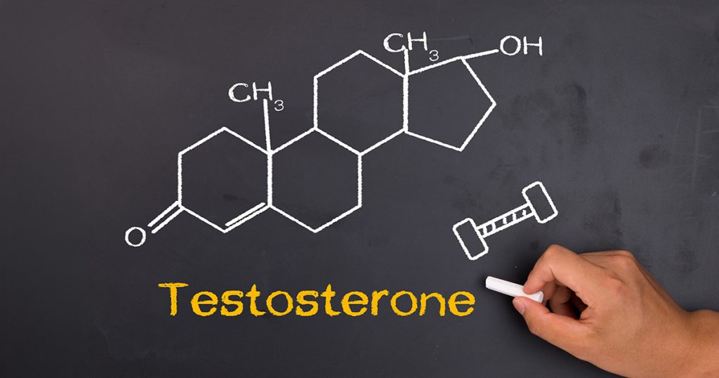
Testosterone biodisponibile negli uomini: fisiopatologia, importanza clinica del desiderio sessuale, implicazioni cardiovascolari e terapia ricostituente con testosterone e hCG
Una revisione clinica per la pratica preventiva basata sulle prove di efficacia
Astratto
La valutazione clinica dello stato androgenico maschile è andata ben oltre la semplice misurazione del siero totale testosterone. Le concentrazioni totali sono abitualmente fuorvianti perché la maggior parte del testosterone circolante è legata al plasma proteine di affinità ampiamente diverse, con la globulina legante gli ormoni sessuali (SHBG) che funge da principale regolatore della quantità di ormone effettivamente rilasciata ai tessuti. Questa rassegna consolida la comprensione moderna di testosterone biodisponibile, che copre la biochimica della compartimentalizzazione, i meccanismi del declino legato all'età, la regolazione della SHBG (comprese le strategie pratiche per abbassarla quando è clinicamente elevata), i metodi analitici che producono misurazioni affidabili e l'importanza clinica del ridotto desiderio sessuale come il segnale clinico più specifico di una vera carenza di androgeni. Vengono esaminate in dettaglio le implicazioni cardiovascolari del basso testosterone biodisponibile, inclusi i risultati dei studio TRAVERSE e l'aggiornamento dell'etichettatura della FDA del 2025. La sezione terapeutica affronta sia la terapia sostitutiva del testosterone sia l'uso appropriato della gonadotropina corionica umana (hCG), che preserva la produzione di testosterone intratesticolare e la spermatogenesi. L'obiettivo è un quadro coerente e contemporaneo che i clinici possano applicare ai pazienti di sesso maschile nella pratica senza confondere il rumore biochimico con la malattia.
1. Introduzione
Questa è la sostanza della ipotesi degli ormoni liberi, e sebbene sia stata perfezionata rispetto alla sua formulazione originaria, la sua affermazione centrale ha retto: il testosterone biodisponibile (la somma delle frazioni libera e legata all'albumina) è la variabile biologica più significativa.1-4
La conseguenza clinica è che due uomini con concentrazioni di testosterone totale identiche possono avere una fisiologia androgenica molto diversa se le loro concentrazioni di SHBG differiscono. La SHBG stessa è regolata da processi epatici che rispondono a insulina, ormoni tiroidei, steroidi sessuali, citochine infiammatorie, stato nutrizionale e invecchiamento. Di conseguenza, condizioni comuni come obesità, di tipo 2 diabete, l'ipertiroidismo, la disfunzione epatica e il processo di invecchiamento possono disaccoppiare il testosterone totale dalla reale attività ormonale nei tessuti.5-7
Questa rassegna è organizzata attorno alle domande a cui un medico deve rispondere nella pratica: perché il testosterone diminuisce con l'età, perché la SHBG aumenta, cosa conta di più dal punto di vista sintomatico (con il desiderio sessuale come indicatore diagnosticamente più utile), come il laboratorio dovrebbe misurare questi analiti, cosa dice ora la letteratura cardiovascolare sia sul testosterone endogeno sia sulla terapia sostitutiva, e come il testosterone e l'hCG debbano essere usati per ripristinare la fisiologia in sicurezza.
2. Compartimentizzazione del testosterone circolante
2.1 Frazioni di legame e loro conseguenze biologiche
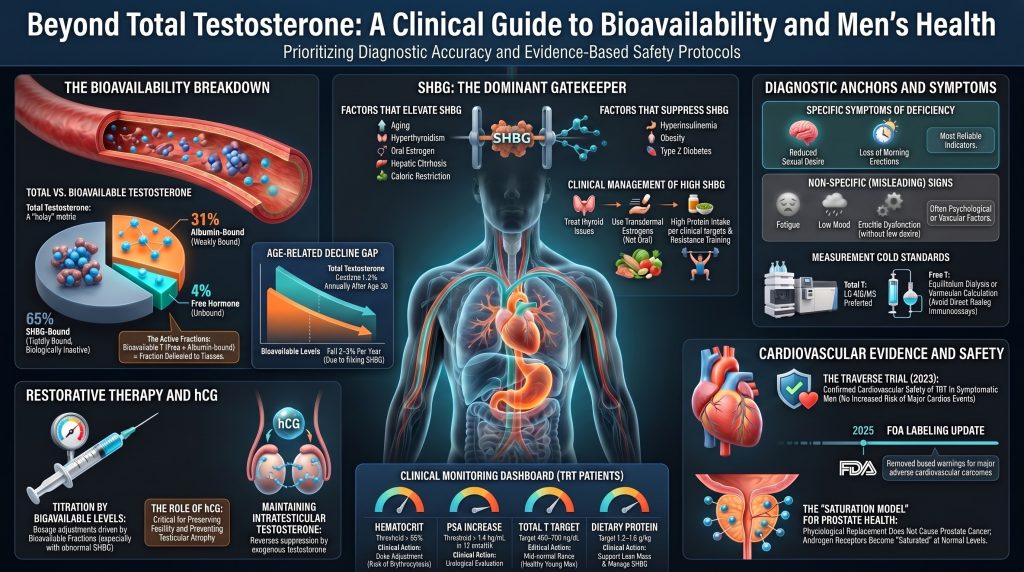
In un uomo sano eugonadico, circa dal 44 al 65 percento del testosterone totale è legato alla SHBG, dal 30 al 50 percento è debolmente legato all'albumina e dall'1 al 4 percento circola come ormone libero.1,2,3 L'SHBG è un omodimero glicoproteico di circa 95 kDa, prodotto principalmente dal fegato. La sua costante di associazione per il testosterone è dell'ordine di 1 × 10⁹ per molare, sufficiente a rendere la frazione legata all'SHBG effettivamente non disponibile durante il breve transito attraverso il letto capillare.1 L'albumina, per contro, è abbondante ma lega il testosterone con un'affinità molto inferiore, pari a circa 3 × 10⁴ per molare.2,3 Il legame con l'albumina si dissocia abbastanza rapidamente che la frazione legata all'albumina può rilasciarsi nei tessuti durante il transito, motivo per cui la somma della frazione libera e di quella legata all'albumina viene definita testosterone biodisponibile.2,4
L'ipotesi dell'ormone libero sostiene che solo la molecola non legata diffonde attraverso la membrana cellulare per agire sul recettore intracellulare degli androgeni.4 Recenti dati in vivo ottenuti con saggi altamente sensibili hanno perfezionato questo quadro, suggerendo che la cinetica di dissociazione della SHBG e dell'albumina consenta una certa biodisponibilità al di là del pool strettamente libero, in particolare nei tessuti con transito capillare lento.2 La conclusione pratica non è cambiata: il solo testosterone totale può trarre in inganno e l'esposizione clinicamente significativa è più vicina alla frazione biodisponibile.
Tabella 1. Frazioni di testosterone plasmatico negli uomini adulti
| Frazione | Partner di legame | Affinità (Ka, M⁻¹) | Quota del totale T | Rilevanza clinica |
| Gratis | Nessuna | Non applicabile | 1–4% | Si diffonde liberamente attraverso le membrane cellulari; frazione attiva |
| Legato all'albumina | Albumina | ~3 × 10⁴ | 30–50% | Debolmente legato; si rilascia durante il transito capillare e contribuisce alla cessione ai tessuti |
| legato alla SHBG | SHBG | ~1 × 10⁹ | 44–65% | Strettamente legato; biologicamente inerte in condizioni normali |
| biodisponibile | Libero + legato all'albumina | Misto | ~30–54% | Miglior indice di esposizione tissutale agli androgeni |
2.2 Perché il testosterone totale non è sufficiente
Poiché le frazioni legate alla SHBG e all’albumina sono regolate da equilibri di azione di massa, una variazione della concentrazione di SHBG modifica la ripartizione tra la frazione legata e quella testosterone libero anche se la produzione testicolare rimane invariata. Un raddoppio dell’SHBG, fenomeno non insolito nel passaggio dalla giovane età adulta alla settima decade di vita, può normalizzare il testosterone totale mentre il testosterone biodisponibile è diminuito in modo sostanziale.2,9,13 Questo è il motivo principale per cui gli uomini più anziani con testosterone totale apparentemente normale possono presentare sintomi clinicamente significativi da carenza di androgeni.
3. Perché il testosterone diminuisce con l'età
Il calo dei livelli di androgeni negli uomini, legato all'età, è ormai ampiamente documentato. Il Studio longitudinale di Baltimora sull’invecchiamento hanno seguito uomini in buona salute per decenni e hanno riportato un calo del testosterone totale pari a circa l'1-2% all'anno dopo i trent'anni, mentre il testosterone libero e quello biodisponibile hanno registrato un calo più marcato, pari a circa il 2-3% all'anno, poiché l'SHBG aumenta parallelamente.9 Il Massachusetts Male Aging Study ha confermato traiettorie simili in una coorte basata sulla comunità.10 Lo studio europeo sull'invecchiamento maschile (EMAS) ha inoltre dimostrato che negli uomini con una normale funzione ipofisaria il declino riflette una combinazione di meccanismi testicolari e centrali.11,15
3.1 Meccanismi di declino
Diverse vie contribuiscono contemporaneamente, ed è per questo che la sindrome di ipogonadismo a esordio tardivo è eterogeneo.
Invecchiamento testicolare. Il numero delle cellule di Leydig diminuisce e quelle sopravvissute Cellule di Leydig mostrano una ridotta capacità steroidogenica. Disfunzione mitocondriale, accumulo di lipofuscina e compromissione della via mediata da StAR colesterolo tutti questi fattori contribuiscono a ridurre la produzione di testosterone per cellula.13
Perdita della pulsatilità ipotalamo-ipofisaria. Gli impulsi di GnRH diventano meno frequenti e di minore ampiezza, gli impulsi di LH fanno altrettanto e l'ampiezza dell'LH necessaria a stimolare la secrezione di testosterone viene raggiunta in modo meno affidabile. Veldhuis e colleghi hanno dimostrato che questa perdita di pulsatilità ordinata è di per sé una componente indipendente dell'invecchiamento riproduttivo maschile.14
Cambiamento della composizione corporea. L'adiposità viscerale si espande durante la mezza età, anche negli uomini che mantengono un peso stabile. Il tessuto adiposo secerne citochine infiammatorie che sopprimono la secrezione di GnRH e gonadotropine ipofisarie e contiene aromatasi, che converte il testosterone in estradiolo. Il risultato è un ipogonadismo secondario funzionale sovrapposto all'invecchiamento testicolare primario.20,21,22
SHBG in aumento. La sintesi epatica di SHBG aumenta di circa l'uno percento all'anno dopo la terza decade nella maggior parte degli uomini.2,9 Poiché la SHBG sequestra il testosterone, a parità di testosterone totale corrisponde una frazione biodisponibile inferiore negli uomini più anziani. Lo studio di armonizzazione di Travison ha stabilito intervalli di riferimento stratificati per età che tengono conto di questa fisiologia.12
Tendenze secolari. Travison e colleghi hanno dimostrato che i livelli di testosterone a livello di popolazione negli uomini americani sono diminuiti tra le coorti di nascita negli ultimi decenni indipendentemente dall'età, suggerendo che i fattori ambientali (probabilmente inclusi la prevalenza dell'obesità, gli interferenti endocrini e i cambiamenti nell'attività fisica) contribuiscono ad aggiungersi al processo di invecchiamento intrinseco.16
3.2 L'effetto dell'invecchiamento sui sintomi
L'EMAS ha stabilito che solo un gruppo ristretto di sintomi traccia in modo affidabile lo stato degli androgeni negli uomini anziani. Tre sintomi sessuali (scarsa erezione mattutina, basso desiderio sessuale e disfunzione erettile) insieme a un testosterone totale inferiore a circa 11 nmol/L (320 ng/dL) e a un testosterone libero inferiore a 220 pmol/L (64 pg/mL) definiscono il fenotipo più specifico dell'ipogonadismo a insorgenza tardiva.11 Gli altri sintomi tipicamente attribuiti al basso livello di testosterone, inclusi stanchezza, umore depresso e forza ridotta, sono comuni negli uomini più anziani ma mostrano una debole correlazione con lo stato androgenico.
4. SHBG: Regolazione e perché è importante
L'SHBG funge da principale regolatore degli androgeni e estrogeno biodisponibilità. La sua sintesi epatica è regolata dall'HNF-4α, con un'azione permissiva o soppressiva da parte dell'insulina, degli ormoni tiroidei, degli steroidi sessuali, delle citochine infiammatorie e dei sensori dei nutrienti.1,5,6,17 È importante sottolineare che un basso livello di SHBG è ormai riconosciuto come un fattore indipendente biomarcatore di insulino-resistenza e il diabete di tipo 2, mentre un livello elevato di SHBG è un indicatore di condizioni epatiche e metaboliche che alterano l'interpretazione di qualsiasi valore di testosterone totale.18,19
4.1 Condizioni che aumentano la SHBG
L'invecchiamento è la causa più comune di elevati livelli di SHBG, con il costante incremento verso l'alto descritto in precedenza. L'ipertiroidismo aumenta la SHBG attraverso la stimolazione di HNF-4α e HNF-1α.17 L'esposizione agli estrogeni (endogeni, da contraccettivi orali o da terapia sostitutiva con estrogeni) induce marcatamente la sintesi epatica della SHBG, che è uno dei meccanismi attraverso cui gli estrogeni orali, ma non transdermici, riducono il testosterone libero nelle donne.6 La cirrosi epatica eleva frequentemente la SHBG (a volte in modo paradossale, data la funzione sintetica epatica altrimenti compromessa) a causa del metabolismo epatocitario alterato e della ridotta clearance.24 L'anoressia nervosa, la grave restrizione calorica e la deprivazione proteica cronica aumentano la SHBG. Diversi anticonvulsivanti (fenitoina, carbamazepina) e alcuni farmaci per l'HIV inducono la SHBG tramite effetti sugli enzimi epatici.6
4.2 Condizioni che abbassano la SHBG
L’iperinsulinemia è la causa clinicamente più rilevante di bassi livelli di SHBG nelle popolazioni odierne. Selva e colleghi hanno dimostrato che i monosaccaridi (in particolare il fruttosio e glucosio) inibiscono la trascrizione dell'SHBG epatico attraverso la sottoregolazione di HNF-4α, con la lipogenesi epatica come mediatore immediato.5 Questo spiega perché un basso livello di SHBG si associ in modo così affidabile a sindrome metabolica, l'obesità e il diabete di tipo 2, e perché l'SHBG viene ora utilizzato come biomarcatore nella ricerca metabolica.18,19,40 Altri soppressori includono l'ipotiroidismo, gli androgeni esogeni (incluso il testosterone sovrafisiologico, gli steroidi anabolizzanti e il danazolo), l'eccesso di glucocorticoidi, l'eccesso di ormone della crescita o IGF-1 e i progestinici con attività androgenica.6
Tabella 2. Principali modulatori della sintesi epatica di SHBG
| Aumenta la SHBG | Abbassa la SHBG |
| Invecchiamento (aumento costante a partire dalla terza decade) | Iperinsulinemia e insulino-resistenza |
| Ipertiroidismo | L'obesità, in particolare l'adiposità viscerale |
| Esposizione a estrogeni orali (contraccettivi orali, gravidanza) | Diabete mellito di tipo 2 |
| Cirrosi epatica | Ipotiroidismo |
| Restrizione calorica e anoressia nervosa | Androgeni esogeni e steroidi anabolizzanti |
| Infezione da HIV (multifattoriale) | Eccesso di glucocorticoidi (sindrome di Cushing) |
| Anticonvulsivanti (fenitoina, carbamazepina) | Acromegalia (eccesso di GH/IGF-1) |
| Cronico infiammazione (effetto variabile) | Sindrome nefrosica (perdita di proteine urinarie) |
4.3 Riduzione della SHBG clinicamente elevata con mezzi non farmacologici
Quando la SHBG è alta in un contesto clinicamente rilevante (comunemente negli uomini anziani con un biotipo corporeo relativamente snello, nell'ipertiroidismo subclinico o negli uomini in terapia con estrogeni per via orale per qualsiasi motivo), la priorità è affrontare la causa sottostante. Le strategie seguenti presentano il maggiore supporto meccanicistico, sebbene la base di prove per molte di esse sia di natura osservazionale piuttosto che randomizzata.
Allenamento con i sovraccarichi e un adeguato apporto proteico nella dieta. Aumenti indotti dall'esercizio fisico in massa magra, in particolare attraverso l'allenamento di resistenza, sono associati a modeste riduzioni dell'SHBG, probabilmente grazie a un miglioramento sensibilità all'insulina a livello muscolare. Un adeguato apporto proteico (da 1,2 a 1,6 g/kg di peso corporeo negli uomini anziani attivi) favorisce l'aumento della massa muscolare e limita lo stato catabolico che, a sua volta, determina un aumento dell'SHBG.20,21
Carboidrati qualità e carico glicemico. L'effetto della modifica della dieta sulla SHBG dipende da quale fattore scatenante predomina e deve essere considerato separatamente per ciascun fenotipo di paziente. Negli uomini insulino-resistenti con SHBG bassa, il miglioramento della sensibilità all'insulina (riducendo zuccheri raffinati, bevande zuccherate con fruttosio e cibi ad alto carico glicemico) tende ad aumentare la SHBG verso la norma. Negli uomini sottonutroiti o con deficit energetico che presentano SHBG alta a causa della restrizione calorica, un adeguato reintegro calorico tende a ridurre la SHBG. Negli uomini con SHBG alta attribuibile a malattie tiroidee, estrogeni orali, anticonvulsivanti o malattie epatiche, la sola modifica della dieta è insufficiente; l'intervento appropriato mira al fattore scatenante sottostante.5,18
Trattamento dell'ipertiroidismo subclinico. Quando il TSH è soppresso e la SHBG è alta, affrontare l'anomalia tiroidea spesso normalizza la SHBG entro pochi mesi.17
Riduzione o sostituzione dell'estrogeno orale. I pazienti in terapia con estrogeni orali per qualsiasi motivo possono passare agli estrogeni transdermici, che bypassano l'effetto di primo passaggio epatico e producono poche variazioni della SHBG.6
Revisione dei farmaci. Gli anticonvulsivanti e determinati antiretrovirali potrebbero richiedere la sostituzione se viene confermato l'ipogonadismo e la SHBG è il fattore principale.6
Affrontare la malattia epatica. Negli uomini affetti da malattia epatica cronica in cui la SHBG risulta paradossalmente elevata, il trattamento del processo epatico (alcol sospensione del trattamento, terapia antivirale per l’epatite C, oppure perdita di peso (per la steatosi epatica) è l'intervento più indicato.24
Adeguato apporto calorico. La SHBG aumenta drasticamente in caso di malnutrizione. Gli atleti e gli uomini anziani in bilancio energetico negativo possono presentare livelli elevati di SHBG che si risolvono con la repletion calorica.6,15
L'integrazione di boro, zinco e magnesio viene talvolta raccomandata per la modulazione della SHBG. Le evidenze cliniche sono limitate e incoerenti, e nessuno di questi sostituisce la risoluzione dei fattori scatenanti a monte sopra menzionati.
5. Condizioni che dissociano il testosterone totale da quello biodisponibile
5.1 Obesità e ipogonadismo secondario funzionale
L'obesità, in particolare l'obesità viscerale, produce un quadro che è stato definito ipogonadismo secondario associato all'obesità maschile (MOSH).22 Questo quadro clinico è caratterizzato da bassi livelli di testosterone totale con livelli di LH e FSH inappropriatamente normali o bassi, il che indica una componente centrale. Tre meccanismi agiscono contemporaneamente: l’iperinsulinemia inibisce la SHBG epatica, riducendo il testosterone totale senza necessariamente abbassare il testosterone libero; il tessuto adiposo viscerale secerne IL-6, il TNF-α e la leptina, secondo meccanismi che inibiscono la secrezione di GnRH da parte dell’ipotalamo; inoltre, l’aromatasi del tessuto adiposo converte il testosterone in estradiolo, fornendo un ulteriore feedback negativo all’ipotalamo.20,22,23 Negli uomini con diabesità, più del 30 percento soddisfa i criteri biochimici per l'ipogonadismo negli studi trasversali.23
Crucialmente, la perdita di peso e il miglioramento della sensibilità all'insulina spesso invertono parzialmente o totalmente questo quadro. La chirurgia bariatrica può normalizzare il testosterone negli uomini obesi ipogonadici, e persino una modesta perdita di peso dal 5 al 10 percento migliora sia il testosterone totale che quello libero.20,21,22
5.2 Malattia epatica steatosica associata a disfunzione metabolica (MASLD)
La steatosi epatica è un'espressione fenotipica dell'insulino-resistenza epatica ed è strettamente associata a bassi livelli di SHBG e di testosterone totale.19,40 Il testosterone libero o biodisponibile è la misurazione più significativa in questi pazienti. La presenza di steatoepatite aggiunge un secondo livello perché la progressione verso la fibrosi e la cirrosi può paradossalmente aumentare la SHBG.24
Cirrosi epatica
La cirrosi avanzata produce un'endocrinopatia complessa. La SHBG aumenta nonostante la ridotta funzione sintetica epatica in molti altri ambiti; l'albumina diminuisce; la clearance dell'estradiolo diminuisce. Il testosterone totale può sembrare ingannevolmente conservato mentre il testosterone libero è crollato.24 La misurazione biodisponibile è obbligatoria in questa popolazione; altrimenti la diagnosi viene mancata.
5.4 Sindrome nefrosica
Una proteinuria massiccia può causare la perdita urinaria di SHBG e albumina, riducendole entrambe. Il testosterone totale diminuisce poiché la capacità di trasporto legata è stata ridotta; il testosterone libero può essere inizialmente preservato dalla compensazione dell'asse HPG, ma lo stress catabolico cronico e gli effetti diretti delle citochine sulle cellule di Leydig finiscono per comprometterne la produzione.25
5.5 Ipertiroidismo e ipotiroidismo
L'ipertiroidismo aumenta la SHBG tramite l'induzione di HNF-1α e HNF-4α, elevando il testosterone totale ma con effetti minori sul testosterone biodisponibile.17 L'ipotiroidismo produce il modello opposto. In qualsiasi paziente in cui il testosterone totale venga interpretato clinicamente, lo stato tiroideo dovrebbe essere noto.
6. Implicazioni cardiovascolari del testosterone biodisponibile
La rilevanza cardiovascolare dello stato del testosterone è la parte di questo campo che è cambiata maggiormente negli ultimi due decenni. La letteratura meno recente considerava il testosterone come cardiovascolarmente neutro o dannoso; le prove attuali supportano una visione più sfumata in cui i livelli fisiologici di testosterone biodisponibile sono associati a un fenotipo cardiovascolare favorevole, mentre sia i livelli molto bassi che quelli soprafisiologici comportano dei rischi.
6.1 Biologia vascolare del testosterone
Il testosterone esercita azioni vascolari dirette. Favorisce la funzione endoteliale ossido nitrico attività sintasica, che favorisce la vasodilatazione endotelio-dipendente. Modula l’attività dei canali del calcio nella muscolatura liscia vascolare, contribuendo alla vasodilatazione coronarica e periferica. Influisce sul tono infiammatorio della parete vascolare; in molte coorti, infatti, bassi livelli di testosterone sono associati a valori elevati di CRP e IL-6.34,50,56 A livello del cardiomiocita, i recettori degli androgeni sono espressi e il supporto androgenico fisiologico appare permissivo per la normale energetica miocardica.
Il testosterone interagisce anche con il lipoproteina sistema in modi clinicamente rilevanti per il rischio di ASCVD. Livelli di testosterone sovrafisiologici, compresi la maggior parte dei casi di abuso di steroidi anabolizzanti, riducono HDL e può aumentare i livelli delle lipoproteine contenenti ApoB. La sostituzione fisiologica, al contrario, produce in genere piccole riduzioni di colesterolo totale e ApoB e lievi riduzioni dell'HDL, con effetti neutri o favorevoli sulla sensibilità all'insulina e grasso viscerale.49,50,56 L'impatto su placca La biologia derivante dal ripristino della fisiologia normale, in contrapposizione all’esposizione sovrafisiologica, è la variabile determinante ai fini della prevenzione.
6.2 Testosterone endogeno ed eventi cardiovascolari
Grande coorti prospettiche È stato generalmente riscontrato che bassi livelli di testosterone endogeno sono associati a un aumento del rischio cardiovascolare e mortalità per tutte le cause negli uomini. L’analisi aggregata condotta da Yeap e colleghi, che ha combinato 11 coorti prospettiche e oltre 24.000 uomini, ha riportato che gli uomini nel quintile più basso di testosterone totale presentavano un rischio maggiore di eventi cardiovascolari e che livelli molto bassi di testosterone libero (inferiori a circa 184 pmol/L, ovvero 53 pg/mL) comportava il rischio maggiore.34 A meta-analisi Corona e colleghi hanno riscontrato associazioni inverse simili tra il testosterone endogeno e gli eventi cardiovascolari.35 Randomizzazione mendeliana Le analisi hanno prodotto risultati più contrastanti, ma in generale non confermano l’esistenza di un effetto causale nocivo del testosterone fisiologico sugli esiti cardiovascolari; alcune analisi suggeriscono invece un’associazione protettiva nella fascia più bassa della distribuzione.55
La stessa SHBG appare essere un biomarcatore cardiovascolare e metabolico indipendente. Bassi livelli di SHBG predicono il diabete di tipo 2 incidente,18 e livelli molto elevati di SHBG negli uomini anziani sono stati associati alla mortalità in alcune coorti, sebbene l’interpretazione sia complicata da causalità inversa (La fragilità e la malnutrizione determinano un aumento della SHBG).19,58
6.3 Sostituzione di testosterone e rischio cardiovascolare: evidenze attuali dopo lo studio TRAVERSE
Nel 2013 e nel 2014, due studi ampiamente pubblicizzati (Vigen et al. nel sistema VA e Finkle et al. in un database di sinistri/rimborsi) hanno riportato associazioni tra la terapia sostitutiva con testosterone (TRT) e gli eventi cardiovascolari. Entrambi presentavano gravi limiti metodologici, tra cui la registrazione incompleta del monitoraggio del testosterone, un breve follow-up e problemi di selezione. Successive analisi hanno sollevato perplessità metodologiche su entrambi gli studi.54, 54b Nonostante ciò, nel 2015 la FDA ha aggiunto un'avvertenza di classe sui possibili rischi cardiovascolari, il che ha ridotto le prescrizioni.
Da allora, numerosi studi osservazionali condotti su popolazioni meglio definite hanno riportato associazioni neutre o benefiche. Sharma e colleghi, in una coorte del Dipartimento degli Affari dei Veterani (VA) composta da oltre 83.000 uomini, hanno riscontrato che la normalizzazione dei livelli di testosterone mediante terapia sostitutiva con testosterone (TRT) era associata a una ridotta incidenza di infarto miocardico, ictus, e la mortalità per tutte le cause rispetto agli uomini i cui livelli sono rimasti bassi o non sono stati trattati.37 Cheetham e colleghi hanno riportato un rischio inferiore del 33% di eventi cardiovascolari negli uomini trattati con TRT nei dati del Kaiser Permanente.38 Anderson e colleghi, all'interno del sistema sanitario Intermountain Healthcare, hanno riportato in modo simile esiti cardiovascolari neutri o favorevoli.39
Il più ampio studio randomizzato sulla sicurezza cardiovascolare condotto finora è stato TRAVERSE (Lincoff et al., New England Journal of Medicine, 2023), uno studio randomizzato, controllato con placebo, di non inferiorità condotto su circa 5.200 uomini di età compresa tra i 45 e gli 80 anni affetti da ipogonadismo e con una delle seguenti condizioni preesistenti malattia cardiovascolare o ad alto rischio cardiovascolare. Nel corso di un periodo di follow-up medio di 22 mesi, l’endpoint primario composito costituito da morte cardiovascolare, infarto miocardico non fatale e ictus non fatale si è verificato nel 7,0% del gruppo trattato con testosterone e nel 7,3% del placebo gruppo, soddisfacendo il criterio prestabilito di non inferiorità (hazard ratio 0,96; intervallo di confidenza 95% (0,78–1,17).36 Fibrillazione atriale, il danno renale acuto e l’embolia polmonare sono risultati leggermente più frequenti nel gruppo trattato con testosterone, in linea con le caratteristiche biologiche note dell’effetto indotto dal testosterone eritrocitosi; questi sono i principali segnali di sicurezza residui che richiedono un monitoraggio. TRAVERSE era uno studio di non inferiorità con una durata del follow-up moderata e non dimostra un beneficio cardiovascolare a lungo termine né affronta in modo esaustivo la biologia della placca, pressione sanguigna, oppure un impiego off-label più ampio.
Nel 2025, la FDA ha emesso modifiche all'etichettatura valide per l'intera classe per i prodotti a base di testosterone. Le etichette aggiornate hanno incorporato i risultati dello studio TRAVERSE e hanno rimosso il linguaggio dell'avvertenza con riquadro nero (boxed warning) riguardante il rischio aumentato di eventi cardiovascolari avversi maggiori. Le etichette riviste hanno aggiunto o rafforzato le avvertenze relative all'innalzamento della pressione sanguigna e hanno mantenuto le limitazioni d'uso esistenti per l'ipogonadismo legato all'età in assenza di una carenza biochimica documentata. I medici dovrebbero consultare le informazioni di prescrizione correnti per la specifica formulazione utilizzata.
6.4 Testosterone e placca coronarica
Lo studio secondario sui rischi cardiovascolari del Testosterone Trials (Budoff et al., JAMA, 2017) ha riportato che gli uomini trattati con gel di testosterone per un anno hanno mostrato un aumento maggiore delle placche coronariche non calcificate volume della placca su angio-TAC coronarica rispetto ai soggetti trattati con placebo.48 Lo studio non era stato progettato per valutare gli eventi clinici, l’aumento di placca non calcificata era modesto e le implicazioni cliniche a lungo termine rimangono incerte. Se considerata insieme allo studio TRAVERSE, l’interpretazione più ragionevole è che non sia stato dimostrato che il ripristino fisiologico aumenti gli eventi cardiovascolari clinici in un periodo di follow-up di durata moderata, mentre gli endpoint relativi all’imaging della placca mostrano segnali che giustificano una sorveglianza continua.
6.5 Eritrocitosi e rischio trombotico
Il segno avverso cardiovascolare più costantemente riproducibile della terapia sostitutiva con testosterone (TRT) è l’eritrocitosi. Il testosterone stimola l’eritropoiesi sia attraverso effetti diretti sul midollo osseo sia attraverso un aumento della produzione di eritropoietina, con un’entità dell’effetto che dipende dalla formulazione. Gli esteri iniettabili producono, al picco, gli incrementi maggiori; i preparati transdermici quelli minori. A ematocrito un valore superiore al 54% è generalmente considerato un'indicazione a ridurre la dose, cambiare la formulazione o sospendere temporaneamente la terapia, nonché a valutare fattori di confondimento come quella ostruttiva apnea notturna e la disidratazione.7,54
6.6 Sintesi per il medico cardiovascolare
Livelli di testosterone endogeno nella parte inferiore dell'intervallo fisiologico sono associati a un aumento del rischio cardiovascolare, il che rappresenta quasi certamente una combinazione di vera vulnerabilità biologica e confondente a causa di un fenotipo metabolico sfavorevole. Non è stato dimostrato che il ripristino dei livelli di testosterone entro i valori fisiologici negli uomini con ipogonadismo sintomatico, sottoposti a un adeguato monitoraggio, aumenti eventi avversi cardiovascolari maggiori nei periodi di follow-up oggetto dello studio. È necessario monitorare l’eritrocitosi. Gestione intensiva delle patologie cardiovascolari convenzionali fattori di rischio (riduzione dei livelli di ApoB, controllo della pressione arteriosa, controllo glicemico, fumo (smettere di fumare e attività fisica) rimane l'intervento cardiovascolare principale. La terapia sostitutiva con testosterone è un trattamento aggiuntivo che mira a colmare una specifica carenza, piuttosto che una terapia cardiovascolare a sé stante.
7. Desiderio Sessuale: Il Sintomo Più Utile per la Diagnosi di Carenza di Testosterone
Di tutti i sintomi attribuiti al basso livello di testosterone, la ridotta libido è quello più affidabilmente e specificamente legato allo stato androgenico. Questo non è un dettaglio di poco conto; è il punto di riferimento diagnostico più utile che un clinico possiede quando si trova a districarsi tra i disturbi aspecifici che spesso accompagnano l'invecchiamento e la malattia metabolica. È importante notare che l'Endocrine Society raccomanda di diagnosticare l'ipogonadismo solo negli uomini che presentano sintomi coerenti con la carenza di testosterone e concentrazioni di testosterone inequivocabilmente e costantemente basse, confermate da almeno due misurazioni mattutine separate; la sola presenza dei sintomi non è sufficiente.7
7.1 Perché il desiderio sessuale ridotto è il sintomo più utile dal punto di vista diagnostico
Travison e colleghi, analizzando il Massachusetts Male Aging Study, hanno dimostrato una relazione graduale tra il testosterone biodisponibile e la libido che si mantiene lungo l'intero intervallo fisiologico, mentre la relazione tra il testosterone e la funzione erettile è molto meno consistente.29 L'EMAS ha identificato indipendentemente tre sintomi sessuali (scarsa erezione mattutina, basso desiderio sessuale e disfunzione erettile) come gli unici sintomi che correlavano con il basso testosterono con ragionevole specificità una volta tenuto conto dell'età.11 Dei tre, il basso desiderio è il più specifico per lo stato androgenico; la disfunzione erettile è fortemente contaminata da cause vascolari, neurogene e farmacologiche, e le erezioni mattutine sono un indicatore sensibile ma meno specifico.
Negli studi randomizzati sulla terapia sostitutiva a base di testosterone, l'effetto più coerente e di maggiore entità riguarda il desiderio sessuale. Lo studio sul sottogruppo della funzione sessuale del Testosterone Trial (Cunningham et al., 2016) ha dimostrato che, dei tre principali esiti sessuali, l'aumento dei punteggi del desiderio è stato sia il più coerente tra gli uomini sia il più clinicamente significativo.30 Le meta-analisi degli effetti della TRT sulla funzione sessuale mostrano analogamente che la libido è il segnale dominante, con effetti più modesti e variabili sulla funzione erettile.32
7.2 Cosa descrivono i pazienti e cosa chiedere
Gli uomini con libido androgeno-carente descrivono un calo fondamentale dell'interesse spontaneo che in precedenza iniziava il pensiero e il comportamento sessuale. Notano di non pensare più al sesso senza sollecitazioni, di non trovare più stimoli ordinari eroticamente rilevanti e spesso descrivono il cambiamento come la perdita di una spinta interna piuttosto che la perdita di capacità. Gli uomini sono solitamente in grado di riferire se la loro libido è cambiata rispetto al proprio livello di riferimento passato, il che è più informativo dal punto di vista diagnostico rispetto alle soglie di riferimento della popolazione.
La perdita delle erezioni notturne e mattutine è strettamente correlata e spesso co-segnalata. L'intervista clinica dovrebbe indagare esplicitamente sia sul desiderio che sulle erezioni mattutine. Chiedere solo della funzione erettile produce risposte che confondono contributi vascolari, neurologici, farmacologici e androgeni; chiedere specificamente del desiderio e delle erezioni mattutine separa questi aspetti.
7.3 Quali sintomi sessuali non predicono
La disfunzione erettile senza calo del desiderio sessuale raramente si risolve con la sola terapia sostitutiva a base di testosterone. Negli uomini con disfunzione erettile isolata e livelli di testosterone biodisponibile inequivocabilmente normali, la diagnosi differenziale è dominata dalle patologie vascolari (spesso la prima manifestazione di una aterosclerosi), cause neurogeniche, antim ipertensivo farmaci, antidepressivi e fattori psicologici. Il trattamento di questi pazienti con testosterone è raramente efficace e li espone agli effetti collaterali della terapia sostitutiva senza che ne derivino benefici corrispondenti.32,33
8. Altre manifestazioni della carenza di androgeni
Oltre ai sintomi sessuali, la sindrome da carenza di testosterone comprende caratteristiche fisiche, metaboliche e psicologiche. Nessuna di queste è specifica quanto la scarsa libido, ma, nel giusto contesto clinico, contribuiscono a confermare la diagnosi.
8.1 Composizione corporea e metabolismo
Gli uomini affetti da ipogonadismo accumulano tessuto adiposo viscerale e perdono massa muscolare magra, con un conseguente calo della forza.20,21,49,60 Ciò è in parte una conseguenza diretta della perdita della segnalazione androgenica a livello del tessuto muscolare e adiposo, e in parte una conseguenza del contesto di sindrome metabolica in cui si sviluppa l’ipogonadismo indotto dall’obesità.59 Studi di sostituzione a lungo termine condotti su uomini obesi affetti da ipogonadismo hanno dimostrato riduzioni significative in circonferenza vita, il peso corporeo e l’HbA1c; tali effetti sono spesso più modesti rispetto a quelli ottenuti con un intervento intensivo sullo stile di vita, ma sembrano aggiungersi a essi.49
8.2 Osso
Il testosterone, in parte attraverso l'aromatizzazione in estradiolo, favorisce la densità minerale ossea negli uomini. Lo studio secondario sulle ossa condotto nell'ambito del Testosterone Trial ha dimostrato aumenti significativi della densità ossea volumetrica e della resistenza ossea stimata negli uomini anziani con bassi livelli di testosterone dopo un anno di terapia sostitutiva.46
8.3 Anemia
L'anemia lieve è comune nell'ipogonadismo non trattato. Lo studio secondario sull'anemia condotto nell'ambito del Testosterone Trial ha evidenziato la correzione dell'anemia in una percentuale significativa di uomini trattati, compresi alcuni nei quali non era possibile individuare una causa alternativa.47
8.4 Umore, motivazione e funzioni cognitive
Molti uomini affetti da ipogonadismo riferiscono stati d’animo depressi, irritabilità e una diminuzione dell’energia; tali sintomi tendono a migliorare in misura modesta con la terapia sostitutiva, sebbene possano avere molte altre cause e non debbano costituire di per sé l’unico elemento su cui basare una diagnosi.7,33 Negli studi randomizzati, gli effetti cognitivi della terapia sostitutiva negli uomini anziani con lievi disturbi della memoria sono risultati modesti e incostanti.57
9. Misurazione: metodi efficaci e metodi fuorvianti
9.1 Testosterone totale
Da oltre un decennio, l’Endocrine Society e il Programma di standardizzazione ormonale del CDC raccomandano l’LC-MS/MS come metodo di riferimento per la misurazione del testosterone totale, in particolare alle concentrazioni più basse rilevanti ai fini diagnostici. I test immunologici basati su piattaforme di vecchia generazione sono affidabili nell’intervallo medio-alto dei valori nei maschi adulti, ma presentano un margine di errore significativo alle basse concentrazioni e non sono raccomandati nelle donne.26,27
Le variabili preanalitiche sono importanti. Il testosterone totale presenta un picco mattutino marcato e un minimo pomeridiano ridotto, con un’ampiezza diurna attenuata negli uomini più anziani. I campioni devono essere prelevati tra le 7 e le 10 del mattino, a digiuno, e in almeno due occasioni distinte prima di confermare una diagnosi di ipogonadismo. Malattie acute, esercizio fisico intenso recente e restrizione calorica recente possono abbassare transitoriamente il testosterone e devono essere presi in considerazione prima di interpretare un valore basso isolato.
9.2 Testosterone libero: metodi, in ordine di importanza
I test immunologici analogici diretti per il testosterone libero sono inaffidabili e vengono espressamente sconsigliati dalle principali società scientifiche di endocrinologia e andrologia.26,27 Possono fornire valori che differiscono di diverse volte dalla frazione libera reale, con errori più elevati proprio nelle popolazioni in cui la misurazione è più importante (uomini con livelli anomali di SHBG).
Il metodo di riferimento è la dialisi di equilibrio seguita da LC-MS/MS. Il campione di siero viene dializzato attraverso una membrana semipermeabile in presenza di un tampone; solo l'ormone libero attraversa la membrana; il testosterone presente nel dializzato viene quindi misurato mediante spettrometria di massa. Si tratta del gold standard, ma è un metodo laborioso e non ampiamente disponibile.3,28
Il testosterone libero calcolato utilizzando la formula di Vermeulen ha mostrato un'eccellente concordanza con la dialisi di equilibrio (coefficiente di correlazione r ≈ 0,99 in confronti condotti in modo adeguato), a condizione che il testosterone totale e l'SHBG siano misurati con piattaforme accurate.3,28 Questo calcolo rappresenta il metodo pratico che la maggior parte dei medici dovrebbe utilizzare; il più recente modello allosterico a più fasi di Goldman et al. migliora la precisione nei valori estremi dell'SHBG, ma non è ancora disponibile su larga scala.2
Tabella 3. Metodi per la misurazione o la stima del testosterone libero
| Metodo | Precisione | Uso pratico | Limitazione |
| Test immunologico analogico diretto | Povero | Non consigliato | Sostanzialmente impreciso sia a livelli bassi che alti di SHBG; gli errori sono più marcati nelle popolazioni in cui la misurazione è più importante |
| Dialisi di equilibrio + LC-MS/MS | Norma di riferimento | Ricerca e casi diagnosticamente complessi | Richiede molto lavoro, è lento, costoso e non è ampiamente disponibile nei laboratori clinici di routine |
| Vermeulen ha calcolato la T libera | Elevato (r ≈ 0,99 rispetto alla dialisi di equilibrio) | Uso clinico di routine | Dipende dai livelli di testosterone totale e di SHBG; meno accurato in caso di valori estremi di SHBG |
| Modello allosterico a più fasi di Goldman | Valori massimi ai livelli estremi dell’SHBG | Casi difficili selezionati | Non è ancora ampiamente implementato nei sistemi di laboratorio clinico di routine |
10. Ripristino del testosterone biodisponibile: farmacologia
10.1 Obiettivi terapeutici
L’obiettivo della terapia sostitutiva è quello di alleviare i sintomi (con il desiderio sessuale come endpoint primario), mantenendo al contempo i parametri biochimici entro i valori fisiologici tipici dei giovani uomini sani. Le linee guida della Endocrine Society indicano come obiettivo un livello di testosterone totale nella fascia media della norma, in genere compreso tra 400 e 700 ng/dL (da 14 a 24 nmol/L), con un corrispondente livello di testosterone libero nella fascia media della norma.7 Negli uomini con livelli elevati di SHBG, i valori target del testosterone totale devono essere interpretati con cautela; in questo gruppo, la titolazione dovrebbe basarsi sul testosterone biodisponibile, non su quello totale.
10.2 Formulazioni
I gel e le soluzioni per uso topico garantiscono livelli sierici costanti che si avvicinano al ritmo circadiano fisiologico. Per questo motivo sono considerati la terapia di prima linea in molte linee guida, pur comportando alcuni svantaggi quali l’applicazione quotidiana, lievi reazioni cutanee e il rischio, modesto ma reale, di trasmissione interpersonale.7
Gli esteri intramuscolari (cipionato o enantato di testosterone) sono economici ed efficaci. Il regime classico da 100 a 200 mg ogni 1-2 settimane produce picchi e valli marcati; molti medici ora preferiscono iniezioni più piccole e più frequenti (ad esempio, da 50 a 80 mg due volte alla settimana, spesso per via sottocutanea) per appiattire il profilo farmacocinetico e ridurre le oscillazioni dell’ematocrito.7,8
Il testosterone undecanoato a lunga durata d’azione, somministrato tramite iniezione intramuscolare profonda ogni 10-14 settimane dopo una fase di carico, garantisce livelli stabili ed è ampiamente utilizzato al di fuori degli Stati Uniti. La formulazione statunitense richiede l’osservazione in ambulatorio a causa del rischio, storicamente raro, di microembolia polmonare da olio.51
I pellet sottocutanei di testosterone, impiantati ogni 3-6 mesi, garantiscono livelli stabili agli uomini che preferiscono una somministrazione meno frequente.8
Il testosterone undecanoato per via orale (assorbito per via linfatica nelle attuali formulazioni statunitensi) e il gel di testosterone per via intranasale rappresentano ulteriori opzioni, ciascuna con specifiche considerazioni relative al monitoraggio.
10.3 Dosaggio in caso di livelli elevati rispetto a quelli bassi di SHBG
Negli uomini con livelli elevati di SHBG (spesso anziani, magri, con ipertiroidismo subclinico o in terapia con anticonvulsivanti) potrebbero essere necessarie dosi più elevate per raggiungere un livello di biodisponibilità soddisfacente, e il testosterone totale potrebbe dover rientrare nella fascia alta della norma. Negli uomini con bassi livelli di SHBG (tipicamente obesi o insulino-resistenti) le dosi standard possono determinare livelli eccessivi di testosterone libero se il dosaggio viene regolato in base al solo testosterone totale, causando acne, irritabilità ed eritrocitosi accelerata. Il dosaggio dovrebbe essere regolato in base al testosterone biodisponibile calcolato in entrambe le popolazioni.
11. Gonadotropina corionica umana: meccanismo d'azione e impiego clinico
La gonadotropina corionica umana (hCG) agisce come un analogo dell’ormone luteinizzante, legandosi al recettore dell’LH sulle cellule di Leydig e stimolando direttamente la produzione intratesticolare di testosterone. Poiché il testosterone esogeno sopprime l'LH e l'FSH ipofisari e interrompe la stimolazione endogena delle cellule di Leydig (con la conseguenza che il testosterone intratesticolare diminuisce e la spermatogenesi viene soppressa), l'hCG svolge due importanti ruoli clinici: come monoterapia in uomini selezionati e come coadiuvante della terapia sostitutiva con testosterone quando è importante preservare la fertilità.
11.1 Il problema del testosterone intratesticolare
La spermatogenesi richiede concentrazioni di testosterone intratesticolare da circa 50 a 100 volte superiori rispetto al testosterone circolante. Il testosterone esogeno, sopprimendo l’LH, elimina lo stimolo alla produzione di testosterone intratesticolare. Coviello e colleghi hanno dimostrato che una dose bassa di hCG (250 UI per via sottocutanea a giorni alterni), somministrata in concomitanza con il testosterone esogeno, mantiene il testosterone intratesticolare entro i valori normali, mentre il testosterone da solo lo riduce di oltre il 90%.41
Hsieh e colleghi hanno successivamente dimostrato che gli uomini sottoposti a terapia sostitutiva con testosterone in associazione a basse dosi di hCG (in genere 500 UI tre volte alla settimana) mantenevano stabili i parametri seminali per tutta la durata del trattamento, mentre quelli che non assumevano hCG sviluppavano, come previsto, oligospermia o azoospermia.42 Ramasamy e altri hanno sintetizzato l’applicazione pratica della terapia combinata per la preservazione della fertilità.43
11.2 Monoterapia con hCG
Nei giovani affetti da ipogonadismo secondario o misto che desiderano preservare la fertilità, la monoterapia con hCG rappresenta una prima scelta ragionevole. Il dosaggio tipico inizia da 1.000 a 1.500 UI per via sottocutanea due o tre volte alla settimana, con titolazione in base alla risposta del testosterone totale, del testosterone libero e dell’estradiolo. Molti uomini raggiungono livelli di testosterone totale nella parte media della norma con la monoterapia. L’estradiolo può aumentare in misura maggiore rispetto alla terapia sostitutiva con testosterone (poiché l’aromatizzazione testicolare è preservata) e, occasionalmente, è giustificato l’uso selettivo di un inibitore dell’aromatasi; tale scelta non dovrebbe tuttavia essere automatica.41,43,44
L'uso dell'hCG in questo contesto è generalmente off-label nella maggior parte dei paesi e dovrebbe essere personalizzato, idealmente con il coinvolgimento di uno specialista in urologia riproduttiva quando la fertilità è un obiettivo concreto. Le prove a sostegno dei protocolli basati sull'hCG derivano principalmente da studi meccanicistici e da piccoli studi osservazionali o prospettici, piuttosto che da studi su larga scala studi clinici randomizzati controllati.
11.3 hCG come terapia aggiuntiva alla terapia sostitutiva con testosterone
Negli uomini in terapia sostitutiva con testosterone che desiderano preservare la fertilità, o che presentano un’atrofia testicolare fastidiosa, al regime terapeutico a base di testosterone viene aggiunta una dose bassa di hCG (in genere 500 UI due o tre volte alla settimana). Questa dose preserva il testosterone intratesticolare e la spermatogenesi senza provocare aumenti eccessivi dei livelli di testosterone totale o di estradiolo.41,42,43 Come nel caso della monoterapia, anche questo uso combinato è generalmente off-label e le prove a sostegno sono in gran parte di natura osservazionale.
11.4 Protocolli di ripresa dopo la sospensione del testosterone
Gli uomini che hanno fatto uso di testosterone esogeno (sia su prescrizione medica che nell’ambito dell’uso di anabolizzanti androgeni) e che desiderano recuperare la funzione endogena e la fertilità sono candidati ai protocolli di riavvio. I componenti includono tipicamente l’hCG per stimolare il recupero delle cellule di Leydig, un modulatore selettivo dei recettori degli estrogeni (clomifene o tamoxifene) per disinibire la secrezione ipofisaria di gonadotropine e, in alcuni casi, FSH ricombinante qualora la spermatogenesi non riesca a riprendersi. Wenker e colleghi hanno riportato tassi di recupero incoraggianti con tali combinazioni in uomini che tentavano di ripristinare la fertilità dopo l’esposizione ad androgeni esogeni.44,45
11.5 Considerazioni pratiche
L’hCG viene somministrata tramite iniezione sottocutanea. Gli effetti collaterali sono solitamente lievi e comprendono reazioni locali nel sito di iniezione, lieve ginecomastia (in caso di aumento dell’estradiolo) e acne. Il costo è sempre stato variabile e in alcune giurisdizioni sono disponibili formulazioni preparate su misura, laddove il controllo di qualità rappresenta una legittima preoccupazione clinica.
12. Monitoraggio della terapia
L’inizio del trattamento e il suo aggiustamento richiedono un monitoraggio sistematico. Il quadro di riferimento riportato di seguito è in linea con le linee guida della Endocrine Society e dell’AUA, modificate alla luce delle evidenze cardiovascolari esaminate in precedenza.7,8
Situazione iniziale. Testosterone totale (LC-MS/MS o test su piattaforma convalidata), SHBG, testosterone libero o biodisponibile calcolato (metodo di Vermeulen), albumina se si deve effettuare il calcolo, LH e FSH (per distinguere l’ipogonadismo primario da quello secondario), prolattina (in casi selezionati), TSH, emocromo completo (con particolare attenzione all’ematocrito), pannello metabolico completo, profilo lipidico a digiuno comprendente l’ApoB, se disponibile, PSA negli uomini di età superiore ai 40 anni ed esame rettale digitale secondo le linee guida dell’AUA.
Tre mesi. Valutazione dei sintomi (con il desiderio sessuale come endpoint primario), ripetizione della misurazione del testosterone totale (effettuata al momento opportuno in base alla formulazione: al livello minimo per gli esteri intramuscolari, da 2 a 8 ore dopo l’applicazione per i gel), emocromo completo e PSA, se del caso. Un ematocrito superiore al 54% comporta una riduzione della dose o un cambio di formulazione.
Sei e dodici mesi. Stesso gruppo di pazienti, con l’aggiunta di una nuova valutazione della composizione corporea e della circonferenza della vita, della risposta lipidica e degli indici glicemici negli uomini affetti da malattie metaboliche.
Successivamente, con cadenza annuale. Valutazione dei sintomi, testosterone totale e libero, ematocrito, PSA negli uomini di età superiore ai 40 anni e rivalutazione completa del rischio cardiovascolare. Un aumento del PSA superiore a 1,4 ng/mL nell’arco di 12 mesi, oppure un valore confermato di PSA superiore a 4,0 ng/mL, giustificano una valutazione urologica.7,8,53
13. Sicurezza: cosa tenere d’occhio e cosa è stato risolto
13.1 Prostata
I dati attuali non confermano la vecchia convinzione secondo cui la terapia sostitutiva con testosterone a dosi fisiologiche causi il cancro alla prostata negli uomini con una salute prostatica di base normale. Il modello di saturazione proposto da Morgentaler e colleghi, in cui la segnalazione del recettore degli androgeni raggiunge la saturazione all’interno dell’intervallo fisiologico, si adatta meglio ai dati disponibili rispetto al modello di stimolazione lineare implicito nelle raccomandazioni precedenti.52,53 Ciò non significa che la terapia sostitutiva sia sicura in caso di carcinoma prostatico non trattato; significa piuttosto che l’iperplasia prostatica benigna senza complicanze e un livello stabile di PSA non costituiscono controindicazioni alla terapia sostitutiva quando è documentato un ipogonadismo sintomatico.
13.2 Eritrocitosi
Come illustrato nella Sezione 6.5, l’eritrocitosi è l’anomalia di laboratorio più comune che limita il dosaggio nella terapia sostitutiva con testosterone (TRT). Nei pazienti di sesso maschile che presentano eritrocitosi persistente nonostante l’assunzione di dosi adeguate, è opportuno valutare la possibile presenza concomitante di apnea ostruttiva del sonno.
13.3 Fertilità
Il testosterone esogeno inibisce in modo affidabile la spermatogenesi. Gli uomini in età riproduttiva devono essere informati in anticipo e a coloro che desiderano preservare la fertilità dovrebbe essere proposta una terapia combinata a base di hCG.41,42,43,45
13.4 Sistema cardiovascolare
Come illustrato in dettaglio nella Sezione 6, lo studio TRAVERSE fornisce prove di livello 1 secondo cui la terapia sostitutiva ormonale negli uomini con ipogonadismo sintomatico, compresi quelli con elevato rischio cardiovascolare, non ha dimostrato di aumentare il rischio di eventi cardiovascolari avversi maggiori nei periodi di follow-up studiati.36 L'aggiornamento delle indicazioni terapeutiche della FDA del 2025 riflette questi dati. L'eritrocitosi, la fibrillazione atriale, la pressione arteriosa e la tromboembolia venosa rimangono segnali da monitorare.
13.5 Altre considerazioni
L'acne, la ginecomastia lieve (in caso di aumento dell'estradiolo) e l'edema sono effetti correlati alla dose e solitamente gestibili. L'apnea notturna può peggiorare a dosi elevate. È opportuno monitorare gli sbalzi d'umore (in genere un miglioramento, occasionalmente irritabilità a livelli sovrafisiologici) insieme ai parametri di laboratorio.
14. Conclusioni
La diagnosi di deficit di testosterone negli uomini è fondamentalmente una questione di identificazione dell’esposizione agli androgeni a livello tissutale, non di misurazione di un valore riportato in un referto di laboratorio. Il testosterone totale è uno strumento di screening, utile nei giovani con una biologia della SHBG sana, ma spesso fuorviante negli uomini più anziani o in qualsiasi paziente affetto da malattie metaboliche. Il testosterone biodisponibile, calcolato in modo affidabile a partire da una misurazione di alta qualità del testosterone totale e da un valore misurato della SHBG, è la variabile che conta.
La SHBG merita di per sé maggiore attenzione clinica di quanta ne riceva solitamente. I suoi livelli aumentano con l’età, in caso di ipertiroidismo, esposizione agli estrogeni, restrizione calorica e disfunzione epatica. Diminuiscono in caso di iperinsulinemia, obesità e uso di steroidi anabolizzanti. Le principali strategie non farmacologiche per ridurre i livelli clinicamente elevati di SHBG consistono nel migliorare la sensibilità all’insulina, correggere le anomalie tiroidee, passare dagli estrogeni per via orale a quelli transdermici ove indicato, garantire un adeguato apporto calorico e proteico e trattare eventuali patologie epatiche sottostanti.
Dal punto di vista clinico, il desiderio sessuale è il sintomo più utile ai fini diagnostici della carenza di androgeni, a condizione che sia accompagnato dalla conferma biochimica di livelli di testosterone costantemente bassi in almeno due misurazioni mattutine. Un uomo con scarso desiderio sessuale e perdita dell’erezione mattutina, in presenza di un livello confermato di testosterone biodisponibile basso, è il paziente che ha maggiori probabilità di trarre beneficio dalla terapia sostitutiva; un uomo con disfunzione erettile isolata e testosterone biodisponibile normale è invece il paziente che ha minori probabilità di trarne beneficio e che, al contrario, necessita con maggiore probabilità di una valutazione cardiovascolare.
Negli ultimi dieci anni l’attenzione si è concentrata sull’aspetto cardiovascolare. Bassi livelli di testosterone endogeno sono correlati a un aumento della mortalità cardiovascolare e totale. Il ripristino dei livelli fisiologici, monitorato per l’eritrocitosi e la pressione arteriosa, non ha dimostrato di aumentare gli eventi cardiovascolari avversi maggiori nel più ampio studio randomizzato disponibile (TRAVERSE) e, in popolazioni selezionate, potrebbe essere associato a una riduzione del rischio secondo i dati osservazionali. L’aggiornamento dell’etichettatura della FDA del 2025 riflette queste evidenze in continua evoluzione. La terapia sostitutiva non sostituisce la riduzione dell’ApoB e gli interventi sullo stile di vita; è un supporto che affronta un fattore specifico che contribuisce al rischio cardiovascolare e metabolico negli uomini che presentano una vera carenza.
Il ripristino terapeutico si ottiene con preparati a base di testosterone scelti in base allo stile di vita del paziente e alle sue esigenze farmacocinetiche. Negli uomini in età riproduttiva, in quelli che non vogliono subire l’atrofia testicolare o in quelli con ipogonadismo secondario che desiderano mantenere la fertilità, l’hCG (da sola o in combinazione con il testosterone) rappresenta la strategia più appropriata. Il compito del medico è quello di adattare il trattamento alle caratteristiche fisiologiche del paziente e di monitorarlo con attenzione.
Ringraziamenti
L’autore dichiara di aver utilizzato un modello linguistico basato sull’intelligenza artificiale (Claude, Anthropic) nella stesura, nell’organizzazione strutturale e nella revisione editoriale del presente manoscritto. Tutti i contenuti scientifici, le interpretazioni cliniche, i riferimenti bibliografici e la sintesi delle ricerche sottostanti rappresentano il lavoro e il giudizio dell’autore. Tutte le fonti citate sono state individuate e verificate in modo indipendente dall’autore. L’IA è stata utilizzata come strumento di scrittura e revisione; non ha contribuito alla sostanza intellettuale o scientifica della revisione. La presente dichiarazione è resa in conformità con gli standard editoriali emergenti relativi alla preparazione di manoscritti assistita dall’IA.
Riferimenti
- Hammond GL. Diverse roles for sex hormone-binding globulin in reproduction. Biol Reprod. 2011;85(3):431-441. doi:10.1095/biolreprod.111.092593
- Goldman AL, Bhasin S, Wu FCW, Krishna M, Matsumoto AM, Jasuja R. A reappraisal of testosterone’s binding in circulation: physiological and clinical implications. Endocr Rev. 2017;38(4):302-324. doi:10.1210/er.2017-00025
- Vermeulen A, Verdonck L, Kaufman JM. A critical evaluation of simple methods for the estimation of free testosterone in serum. J Clin Endocrinol Metab. 1999;84(10):3666-3672. doi:10.1210/jcem.84.10.6079
- Mendel CM. The free hormone hypothesis: a physiologically based mathematical model. Endocr Rev. 1989;10(3):232-274. doi:10.1210/edrv-10-3-232
- Selva DM, Hogeveen KN, Innis SM, Hammond GL. Monosaccharide-induced lipogenesis regulates the human hepatic sex hormone-binding globulin gene. J Clin Invest. 2007;117(12):3979-3987. doi:10.1172/JCI32249
- Pugeat M, Nader N, Hogeveen K, Raverot G, Déchaud H, Grenot C. Sex hormone-binding globulin gene expression in the liver: drugs and the metabolic syndrome. Mol Cell Endocrinol. 2010;316(1):53-59. doi:10.1016/j.mce.2009.09.020
- Bhasin S, Brito JP, Cunningham GR, et al. Testosterone therapy in men with hypogonadism: an Endocrine Society clinical practice guideline. J Clin Endocrinol Metab. 2018;103(5):1715-1744. doi:10.1210/jc.2018-00229
- Mulhall JP, Trost LW, Brannigan RE, et al. Evaluation and management of testosterone deficiency: AUA guideline. J Urol. 2018;200(2):423-432. doi:10.1016/j.juro.2018.03.115
- Harman SM, Metter EJ, Tobin JD, Pearson J, Blackman MR. Longitudinal effects of aging on serum total and free testosterone levels in healthy men: Baltimore Longitudinal Study of Aging. J Clin Endocrinol Metab. 2001;86(2):724-731. doi:10.1210/jcem.86.2.7219
- Feldman HA, Longcope C, Derby CA, et al. Age trends in the level of serum testosterone and other hormones in middle-aged men: longitudinal results from the Massachusetts Male Aging Study. J Clin Endocrinol Metab. 2002;87(2):589-598. doi:10.1210/jcem.87.2.8201
- Wu FCW, Tajar A, Beynon JM, et al. Identification of late-onset hypogonadism in middle-aged and elderly men. N Engl J Med. 2010;363(2):123-135. doi:10.1056/NEJMoa0911101
- Travison TG, Vesper HW, Orwoll E, et al. Harmonized reference ranges for circulating testosterone levels in men of four cohort studies in the United States and Europe. J Clin Endocrinol Metab. 2017;102(4):1161-1173. doi:10.1210/jc.2016-2935
- Kaufman JM, Vermeulen A. The decline of androgen levels in elderly men and its clinical and therapeutic implications. Endocr Rev. 2005;26(6):833-876. doi:10.1210/er.2004-0013
- Veldhuis JD, Keenan DM, Liu PY, Iranmanesh A, Takahashi PY, Nehra AX. The aging male hypothalamic-pituitary-gonadal axis: pulsatility and feedback. Mol Cell Endocrinol. 2009;299(1):14-22. doi:10.1016/j.mce.2008.09.005
- Camacho EM, Huhtaniemi IT, O’Neill TW, et al. Age-associated changes in hypothalamic-pituitary-testicular function in middle-aged and older men are modified by weight change and lifestyle factors: longitudinal results from the European Male Ageing Study. Eur J Endocrinol. 2013;168(3):445-455. doi:10.1530/EJE-12-0890
- Travison TG, Araujo AB, O’Donnell AB, Kupelian V, McKinlay JB. A population-level decline in serum testosterone levels in American men. J Clin Endocrinol Metab. 2007;92(1):196-202. doi:10.1210/jc.2006-1375
- Selva DM, Hammond GL. Thyroid hormones act indirectly to increase sex hormone-binding globulin production by liver via hepatocyte nuclear factor-4alpha. J Mol Endocrinol. 2009;43(1):19-27. doi:10.1677/JME-09-0025
- Ding EL, Song Y, Manson JE, et al. Sex hormone-binding globulin and risk of type 2 diabetes in women and men. N Engl J Med. 2009;361(12):1152-1163. doi:10.1056/NEJMoa0804381
- Brand JS, van der Tweel I, Grobbee DE, Emmelot-Vonk MH, van der Schouw YT. Testosterone, sex hormone-binding globulin and the metabolic syndrome: a systematic review and meta-analysis of observational studies. Int J Epidemiol. 2011;40(1):189-207. doi:10.1093/ije/dyq158
- Kelly DM, Jones TH. Testosterone and obesity. Obes Rev. 2015;16(7):581-606. doi:10.1111/obr.12282
- Grossmann M. Hypogonadism and male obesity: focus on unresolved questions. Clin Endocrinol (Oxf). 2018;89(1):11-21. doi:10.1111/cen.13723
- Saboor Aftab SA, Kumar S, Barber TM. The role of obesity and type 2 diabetes mellitus in the development of male obesity-associated secondary hypogonadism. Clin Endocrinol (Oxf). 2013;78(3):330-337. doi:10.1111/cen.12092
- Dhindsa S, Ghanim H, Batra M, et al. Hypogonadotropic hypogonadism in men with diabesity. Diabetes Care. 2018;41(7):1516-1525. doi:10.2337/dc17-2510
- Sinclair M, Grossmann M, Gow PJ, Angus PW. Testosterone in men with advanced liver disease: abnormalities and implications. J Gastroenterol Hepatol. 2015;30(2):244-251. doi:10.1111/jgh.12695
- Schiavi A, Carugno M, Tozzi V, et al. Endocrine dysfunction in nephrotic syndrome: a comprehensive review. Biomedicines. 2024;12(8):1860. doi:10.3390/biomedicines12081860
- Rosner W, Auchus RJ, Azziz R, Sluss PM, Raff H. Position statement: utility, limitations, and pitfalls in measuring testosterone: an Endocrine Society position statement. J Clin Endocrinol Metab. 2007;92(2):405-413. doi:10.1210/jc.2006-1864
- Rosner W, Vesper H. Toward excellence in testosterone testing: a consensus statement. J Clin Endocrinol Metab. 2010;95(10):4542-4548. doi:10.1210/jc.2010-1314
- Fiers T, Wu F, Moghetti P, Vanderschueren D, Lapauw B, Kaufman JM. Reassessing free-testosterone calculation by liquid chromatography-tandem mass spectrometry direct equilibrium dialysis. J Clin Endocrinol Metab. 2018;103(6):2167-2174. doi:10.1210/jc.2017-02360
- Travison TG, Morley JE, Araujo AB, O’Donnell AB, McKinlay JB. The relationship between libido and testosterone levels in aging men. J Clin Endocrinol Metab. 2006;91(7):2509-2513. doi:10.1210/jc.2005-2508
- Cunningham GR, Stephens-Shields AJ, Rosen RC, et al. Testosterone treatment and sexual function in older men with low testosterone levels. J Clin Endocrinol Metab. 2016;101(8):3096-3104. doi:10.1210/jc.2016-1645
- Snyder PJ, Bhasin S, Cunningham GR, et al. Effects of testosterone treatment in older men. N Engl J Med. 2016;374(7):611-624. doi:10.1056/NEJMoa1506119
- Corona G, Isidori AM, Buvat J, et al. Testosterone supplementation and sexual function: a meta-analysis study. J Sex Med. 2014;11(6):1577-1592. doi:10.1111/jsm.12536
- Buvat J, Maggi M, Gooren L, et al. Endocrine aspects of male sexual dysfunctions. J Sex Med. 2010;7(4 Pt 2):1627-1656. doi:10.1111/j.1743-6109.2010.01780.x
- Yeap BB, Marriott RJ, Antonio L, et al. Associations of serum testosterone and sex hormone-binding globulin with incident cardiovascular events in middle-aged to older men. Ann Intern Med. 2022;175(2):159-170. doi:10.7326/M21-0551
- Corona G, Rastrelli G, Di Pasquale G, Sforza A, Mannucci E, Maggi M. Endogenous testosterone levels and cardiovascular risk: meta-analysis of observational studies. J Sex Med. 2018;15(9):1260-1271. doi:10.1016/j.jsxm.2018.06.012
- Lincoff AM, Bhasin S, Flevaris P, et al. Cardiovascular safety of testosterone-replacement therapy. N Engl J Med. 2023;389(2):107-117. doi:10.1056/NEJMoa2215025
- Sharma R, Oni OA, Gupta K, et al. Normalization of testosterone level is associated with reduced incidence of myocardial infarction and mortality in men. Eur Heart J. 2015;36(40):2706-2715. doi:10.1093/eurheartj/ehv346
- Cheetham TC, An J, Jacobsen SJ, et al. Association of testosterone replacement with cardiovascular outcomes among men with androgen deficiency. JAMA Intern Med. 2017;177(4):491-499. doi:10.1001/jamainternmed.2016.9546
- Anderson JL, May HT, Lappe DL, et al. Impact of testosterone replacement therapy on myocardial infarction, stroke, and death in men with low testosterone concentrations in an integrated health care system. Am J Cardiol. 2016;117(5):794-799. doi:10.1016/j.amjcard.2015.11.063
- Mohammed M, Al-Habori M, Abdullateef A, Saif-Ali R. Impact of metabolic syndrome factors on testosterone and SHBG in type 2 diabetes mellitus and metabolic syndrome. J Diabetes Res. 2018;2018:4926789. doi:10.1155/2018/4926789
- Coviello AD, Matsumoto AM, Bremner WJ, et al. Low-dose human chorionic gonadotropin maintains intratesticular testosterone in normal men with testosterone-induced gonadotropin suppression. J Clin Endocrinol Metab. 2005;90(5):2595-2602. doi:10.1210/jc.2004-0802
- Hsieh TC, Pastuszak AW, Hwang K, Lipshultz LI. Concomitant intramuscular human chorionic gonadotropin preserves spermatogenesis in men undergoing testosterone replacement therapy. J Urol. 2013;189(2):647-650. doi:10.1016/j.juro.2012.09.043
- Ramasamy R, Armstrong JM, Lipshultz LI. Preserving fertility in the hypogonadal patient: an update. Asian J Androl. 2015;17(2):197-200. doi:10.4103/1008-682X.142772
- Wenker EP, Dupree JM, Langille GM, et al. The use of HCG-based combination therapy for recovery of spermatogenesis after testosterone use. J Sex Med. 2015;12(6):1334-1337. doi:10.1111/jsm.12890
- Crosnoe LE, Grober E, Ohl D, Kim ED. Exogenous testosterone: a preventable cause of male infertility. Transl Androl Urol. 2013;2(2):106-113. doi:10.3978/j.issn.2223-4683.2013.06.01
- Snyder PJ, Kopperdahl DL, Stephens-Shields AJ, et al. Effect of testosterone treatment on volumetric bone density and strength in older men with low testosterone: a controlled clinical trial. JAMA Intern Med. 2017;177(4):471-479. doi:10.1001/jamainternmed.2016.9539
- Roy CN, Snyder PJ, Stephens-Shields AJ, et al. Association of testosterone levels with anemia in older men: a controlled clinical trial. JAMA Intern Med. 2017;177(4):480-490. doi:10.1001/jamainternmed.2016.9540
- Budoff MJ, Ellenberg SS, Lewis CE, et al. Testosterone treatment and coronary artery plaque volume in older men with low testosterone. JAMA. 2017;317(7):708-716. doi:10.1001/jama.2016.21043
- Saad F, Yassin A, Doros G, Haider A. Effects of long-term treatment with testosterone on weight and waist size in 411 hypogonadal men with obesity classes I-III: observational data from two registry studies. Int J Obes (Lond). 2016;40(1):162-170. doi:10.1038/ijo.2015.139
- Traish AM. Benefits and health implications of testosterone therapy in men with testosterone deficiency. Sex Med Rev. 2018;6(1):86-105. doi:10.1016/j.sxmr.2017.10.001
- Hackett G, Cole N, Bhartia M, Kennedy D, Raju J, Wilkinson P. Testosterone replacement therapy with long-acting testosterone undecanoate improves sexual function and quality-of-life parameters versus placebo in a population of men with type 2 diabetes. J Sex Med. 2013;10(6):1612-1627. doi:10.1111/jsm.12146
- Morgentaler A, Zitzmann M, Traish AM, et al. Fundamental concepts regarding testosterone deficiency and treatment: international expert consensus resolutions. Mayo Clin Proc. 2016;91(7):881-896. doi:10.1016/j.mayocp.2016.04.007
- Khera M, Crawford D, Morales A, Salonia A, Morgentaler A. A new era of testosterone and prostate cancer: from physiology to clinical implications. Eur Urol. 2014;65(1):115-123. doi:10.1016/j.eururo.2013.08.015
- Glueck CJ, Wang P. Testosterone therapy, thrombosis, thrombophilia, cardiovascular events. Metabolism. 2014;63(8):989-994. doi:10.1016/j.metabol.2014.05.005
- Mohammadi-Shemirani P, Chong M, Pigeyre M, et al. Effects of lifelong testosterone exposure on health and disease using Mendelian randomization. eLife. 2020;9:e58914. doi:10.7554/eLife.58914
- Mohler ER 3rd, Ellenberg SS, Lewis CE, et al. The effect of testosterone on cardiovascular biomarkers in the testosterone trials. J Clin Endocrinol Metab. 2018;103(2):681-688. doi:10.1210/jc.2017-02243
- Resnick SM, Matsumoto AM, Stephens-Shields AJ, et al. Testosterone treatment and cognitive function in older men with low testosterone and age-associated memory impairment. JAMA. 2017;317(7):717-727. doi:10.1001/jama.2016.20828
- Yeap BB, Knuiman MW, Divitini ML, et al. Differential associations of testosterone, dihydrotestosterone and oestradiol with physical, metabolic and health-related factors in community-dwelling men aged 17-97 years from the Busselton Health Survey. Clin Endocrinol (Oxf). 2014;81(1):100-108. doi:10.1111/cen.12407
- Kupelian V, Page ST, Araujo AB, Travison TG, Bremner WJ, McKinlay JB. Low sex hormone-binding globulin, total testosterone, and symptomatic androgen deficiency are associated with development of the metabolic syndrome in nonobese men. J Clin Endocrinol Metab. 2006;91(3):843-850. doi:10.1210/jc.2005-1326
- Allan CA, Strauss BJ, Burger HG, Forbes EA, McLachlan RI. Testosterone therapy prevents gain in visceral adipose tissue and loss of skeletal muscle in nonobese aging men. J Clin Endocrinol Metab. 2008;93(1):139-146. doi:10.1210/jc.2007-1291

