
Moleculair Verkeer en Arteriële Ziekte
Hoe ApoB-lipoproteïnen en Rho GEF-signaleringsroutes helpen bij het verklaren van atherosclerose
Aderverkalking wordt vaak te simpel uitgelegd. Aan de meeste mensen wordt verteld dat “hoog cholesterol”verstopt de slagaders, en hoewel dat idee in de juiste richting wijst, laat het de diepere biologie achterwege die de ziekte daadwerkelijk veroorzaakt. Cholesterol op zich is niet het hele verhaal. Het nauwkeurigere beeld is dat atherosclerose zich ontwikkelt wanneer atherogene lipoproteïne deeltjes komen het slagader wand, verstrikt raken en een lange ontstekingscascade in werking stellen. Of dat gebeurt, hangt niet alleen af van hoeveel deeltjes er in het bloed circuleren, maar ook van hoe de vaatwand zich op moleculair niveau gedraagt [1-8].
Dat tweede deel is belangrijk-er dan de meeste discussies erkennen. De vaatwand is geen passieve buis. Het is een actief, reagerend weefsel. Endotheelcellen reguleren welke deeltjes door de vaatwand dringen. Immuuncellen beslissen hoe ze reageren zodra die deeltjes zijn vastgehouden. Gladde spiercellen helpt dan bepalen of een tandplak stabiliseert of gevaarlijk wordt. Veel van deze lokale regulering wordt gecontroleerd door een familie van moleculaire schakelaars die bekend staat als Rho-guaninenucleotide-uitwisselingsfactoren, of Rho GEF's, werkt door Rho-GTPasen zoals RhoA, Rac1 en Cdc42 [4].
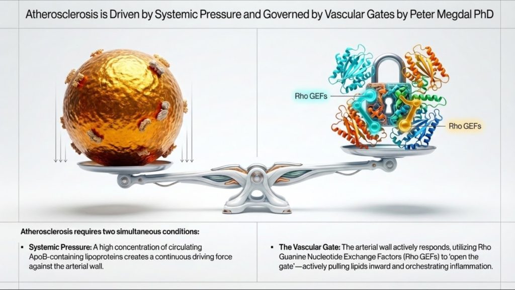
Dit biedt een completere manier om na te denken over hart- en vaatziekten. Systemische lipidenbelasting, met name het aantal ApoB-bevattende deeltjes, bepaalt de druk in het systeem. Maar lokale signaaloverdracht binnen de vaatwand bepaalt of die deeltjes worden toegelaten, vastgehouden en getransformeerd in plaque. In praktijktermen, ApoB meet het aantal voertuigen op de weg, terwijl de Rho GEF-signaaloverdracht helpt te bepalen hoe open de poort is en hoe destructief de lokale reactie zal zijn1-4].
Het begrijpen van dit verband helpt om de kloof te overbruggen tussen klinische lipidologie op hoog niveau en de moderne vasculaire biologie. Het maakt de ziekte ook gemakkelijker uit te leggen aan het grote publiek. Aderverkalking is niet alleen “vet in het bloed”. Het is een probleem van deeltjesverkeer, arteriële toetreding, retentie, immuunactivatie en weefselremodeling.
Waarom Lipoproteïnen überhaupt bestaan
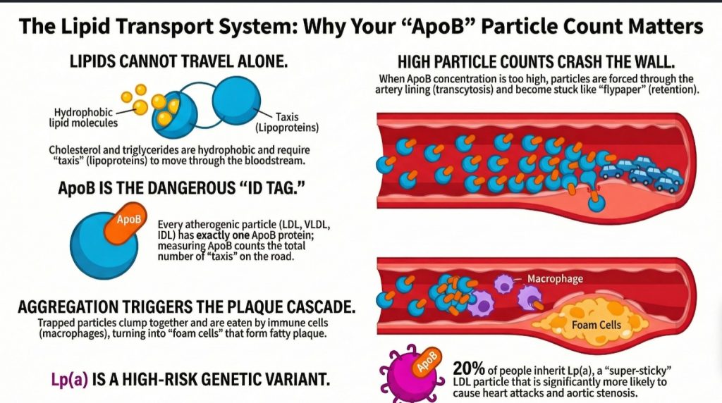
Het menselijk lichaam heeft een fundamenteel technisch probleem. Het moet lipiden transporteren zoals triglyceriden en cholesterol, maar bloedplasma bestaat grotendeels uit water. Lipiden zijn hydrofoob, wat betekent dat ze niet goed oplossen in een watermilieu. Als het lichaam zomaar onbewerkte vetten in de bloedbaan zou afgeven, zouden deze zich afscheiden en samenklonteren, net zoals olie in water [1,2].
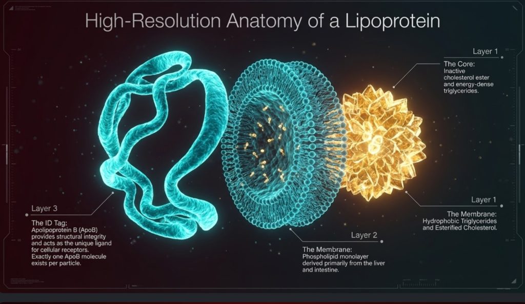
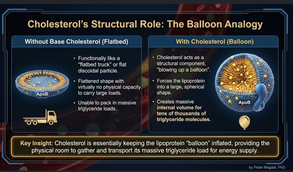
De oplossing is het lipoproteïne. Lipoproteïnen zijn transportdeeltjes die zijn gebouwd om hydrofobe lading door waterachtig plasma te vervoeren. Ze hebben een hydrofobe kern, waar triglyceriden en cholesterolesters worden opgeslagen, en een beter met water compatibel oppervlak dat is gemaakt van fosfolipiden, vrij cholesterol en eiwitten gebeld apolipoproteïnen [1,2,5].
Dit is belangrijk omdat lipoproteïnen vaak worden behandeld als abstracte laboratoriumwaarden, maar ze beter kunnen worden begrepen als fysieke transportvoertuigen. Ze verplaatsen energie en bouwstoffen door het lichaam. Triglyceriden zijn de belangrijkste energielading. Cholesterol zorgt voor structurele ondersteuning van membranen, de synthese van steroïadhormonen en galzuur productie. De bloedbaan is de snelweg, en lipoproteïnen zijn de vloot1-3,5].
Vanuit dat oogpunt is het lichaam lipoproteins niet hoofdzakelijk gaan evolueren om cholesterol te verplaatsen. Het evolueerde ze om energie te verplaatsen. Cholesterol is noodzakelijk, maar het transportsysteem is diep verbonden met brandstoftoevoer. Dat onderscheid is belangrijk, omdat het helpt om een van de meest misverstane kwesties bij hart- en vaatziekten te verhelderen: het lichaam heeft cholesterol weliswaar nodig, maar het heeft geen grote hoeveelheden cholesterol nodig die door het plasma bewegen om weefsels in leven en functionerend te houden [2,11-15].
Cholesterolhomeostase: Hoe weinig er werkelijk nodig is
Een centraal maar vaak over het hoofd gezien feit in de lipidenbiologie is dat het lichaam op cellulair niveau slechts een zeer kleine hoeveelheid cholesterol nodig heeft. Elke celmembraan heeft cholesterol nodig voor structuur en vloeibaarheid, en bepaalde gespecialiseerde weeën hebben het nodig voor de synthese van steroïd hormonen. Maar die behoefte is opmerkelijk klein, streng gereguleerd en wordt grotendeels lokaal bevredigd in plaats van via massale aanvoer vanuit circulerende LDL [2,11-17].
Cholesterolhomeostase wordt gereguleerd door strikt gereguleerde intracellulaire signaalroutes die zorgen voor voldoende aanvoer en tegelijkertijd toxiciteit voorkomen. In tegenstelling tot wat vaak wordt aangenomen, bezitten vrijwel alle celkernhoudende cellen het vermogen om hun eigen cholesterol te synthetiseren. Ze wachten niet simpelweg tot lipoproteïnen het aanleveren. Ze zijn genetisch uitgerust om te maken wat ze nodig hebben [11,12].
Het centrale regelgevende systeem is SREBP-2-SCAP-INSIG-as. Wanneer de intracellulaire cholesterolspiegels dalen, sterol regulatoire element-bindend eiwit 2 (SREBP-2) wordt begeleid door SREBP-splitsingsactiverend eiwit (SCAP) van het endoplasmatisch reticulum naar het Golgi-apparaat. Daar ondergaat SREBP-2 proteolytische activatie. De actieve transcriptiefactor gaat vervolgens de celkern binnen en reguleert genen op die betrokken zijn bij cholesterolsynthese en opname, inclusief HMG-CoA-reductase (HMGCR) en de LDL-receptor (LDLR) [11,12].
Dit stuurt de mevalonaatroute, beginnend met acetyl-CoA en verder gaand via HMG-CoA en mevalonaat richting de cholesterolsynthese. Omdat dit mechanisme door het hele lichaam aanwezig is, zijn cellen niet afhankelijk van grote plasma-pools van cholesterol om de membraanintegriteit of het basisoverleven te waarborgen [11,12].
Wanneer het intracellulaire cholesterol stijgt, schakelt het systeem zichzelf uit. Cholesterol en oxysterolen bevorderen INSIG-gemedieerde retentie van het SCAP-SREBP-complex in het endoplasmatisch reticulum, waardoor verdere transcriptionele activatie wordt verminderd. Tegelijkertijd, HMG-CoA-reductase is bestemd voor afbraak via ubiquitine-proteasoom-routes. Met andere woorden, het cholesterolmetabolisme wordt gereguleerd door een ingebouwde terugkoppelingslus die is ontworpen om buitensporige ophoping te voorkomen [11,12].
Dat overschot is van belang, omdat vrij cholesterol niet goedaardig is wanneer het zich ophopt in cellen. Te veel intracellulair cholesterol kan membranen destabiliseren, kristalliseren in lysosomen en ontstekingssignalering opwekken zoals de NLRP3-inflammasoom, en bevorderen apoptose of necrose13,14Dit is één reden waarom het lichaam er niet op is ontworpen om het op te slaan.
Cellen vertrouwen daarom niet alleen op synthese en feedbackremming, maar ook op actieve export. Door Lever X-receptor (LXR) signalering, oxysterolen induceren transporters zoals ABCA1 en ABCG1, die overtollige cholesterol uit cellen wegvoeren en naar HDL-gemedieerde omgekeerd cholesteroltransport [15].
Samen laten deze routes iets belangrijk zien: cellen hebben cholesterol nodig, maar alleen in kleine, gecontroleerde hoeveelheden. Het voornaamste biologische probleem is niet het verkrijgen van meer cholesterol. Het is het in evenwicht brengen van de synthese, het gebruik, de opslag en de efflux zodat een teveel niet giftig wordt11-15].
Weefselspecifieke cholesterolonafhankelijkheid
Dit principe wordt nog duidelijker wanneer je kijkt naar specifieke organen. Veel weefsels synthetiseren lokaal cholesterol en zijn niet sterk afhankelijk van circulerend LDL.
De centraal zenuwstelsel is het meest treffende voorbeeld. Omdat apoB-bevattende lipoproteïnen de bloed-hersenbarrière niet betekenisvol passeren, moet de hersenstructuur grotendeels zijn eigen cholesterol produceren. Astrocyten cholesterol synthetiseren en het lokaal distribueren via apoE-bevattende lipoproteïnen, een proces dat is gekoppeld aan LXR-gereguleerde routes [16].
Hetzelfde geldt in grote lijnen voor steroïdogene weefsels, met inbegrip van de bijnieren en gonaden. Deze weefsels kunnen cholesterol de novo synthetiseren uit acetaat. Hoewel ze receptoren tot expressie brengen zoals LDLR en SR-B1, hun afhankelijkheid van circulerend LDL onder gewone omstandigheden lijkt beperkt te zijn, waarbij de opname belangrijker wordt in gespecialiseerde of veeleisende toestanden zoals acute ACTH-stimulatie [2,17].
Dit ondersteunt een belangrijk fysiologisch punt. Het lichaam is niet afhankelijk van hoge circulerende LDL-C-waarden voor een normale endocriene of cellulaire functie. Inderdaad hebben mechanistische en klinische discussies in de lipidologie benadrukt dat zeer lage LDL-C-waarden, zelfs rond 10 mg/dL, impliceren niet noodzakelijkerwijs verminderde steroïdogenese of membraanfalen wanneer de intracellulaire synthese intact blijft [2,11,17].
Dus hoewel cholesterol essentieel is, is de hoeveelheid die daadwerkelijk in de bloedbaan nodig is voor celoverleving veel kleiner dan veel mensen aannemen. Die realiteit versterkt de visie dat verhoogde circulerende ApoB-partikels geen biologische noodzaak zijn; het is een blootstelling aan risico.
De ApoB-vloot: Waarom het aantal deeltjes ertoe doet
Van de vele circulerende lipoproteïnen zijn de ApoB-bevattend deeltjes het meest relevant voor atherosclerose. Deze omvatten chylomicronen en hun remnanten, zeer lage-dichtheid-lipoproteïnen (VLDL), IDL (intermediaire-dichtheid lipoproteïnen (IDL), lage-dichtheidlipoproteïnen (LDL), en lipoproteïne(a), of Lp(a) [1,2].
Het belangrijkste klinische inzicht is dat elk atherogeen deeltje bevat precies één ApoB-molecuul. Dat betekent dat ApoB dient als een directe telling van het aantal atherogene deeltjes in de circulatie. Daarentegen meet LDL-C alleen de massa van de cholesterol in LDL-deeltjes. Een persoon kan een relatief normaal LDL-C hebben, maar toch een hoog aantal deeltjes hebben als die deeltjes klein en cholesterolarm zijn. In dat opzicht kan het risico worden onderschat als clinici alleen naar LDL-C kijken [1-3].
Dit is waarom veel lipidendeskundigen ApoB inmiddels beschouwen als de biologisch meest zinvolle maatstaf. Het weerspiegelt hoeveel kansen de deeltjes hebben om de vaatwand binnen te dringen. Simpel gezegd: als elk ApoB-deeltje één potentiële arteriële botsing is, vertelt ApoB ons de verkeersdichtheid.1-3].
Die verkeersanalogie wordt met name nuttig bij het uitleggen van risico's aan niet-specialisten. Het probleem is niet alleen hoeveel vracht er in elke vrachtwagen zit. Het probleem is hoeveel vrachtwagens er op de weg zijn, hoe lang ze daar blijven, en hoe groot de kans is dat ze tegen de zijmuur botsen.
Dit is ook het punt waarop de discussie over minimaal cholesterol klinisch relevant wordt. Als weefsels de bescheiden hoeveelheid cholesterol die ze nodig hebben zelf kunnen synthetiseren, dan kunnen hoge ApoB-waarden niet het best worden opgevat als een nuttige leveringsreserve. Ze kunnen beter worden begrepen als een verhoogde belasting van potentieel deeltjes die in de slagader doordringen2,11-17].
Van Brandstoflevering tot LDL-persistentie
Het verhaal begint met triglyceriderijke deeltjes. De darm produceert chylomicronen om dieetvet te transporteren. De lever produceert VLDL om endogene triglyceriden te transporteren. Dit zijn grote deeltjes waarvan de belangrijkste taak het leveren van brandstof is [1,2].
Terwijl deze deeltjes circuleren, interageren ze met lipoproteïnelipase (LPL), een enzym dat triglyceriden loskoppelt voor gebruik door spier-, hart- en vetweefsel. Naarmate triglyceriden worden verwijderd, krimpt het deeltje. Oppervlaktemateriaal komt vrij. Maar de ApoB-ruggengraat blijft achter. Na verloop van tijd wordt VLDL omgevormd tot IDL en vervolgens tot LDL [1,2,18].
LDL is daarom niet van nature een of andere schurkachtige of kwaadaardige deeltje. Het is een stroomafwaarts product van het normale lipoproteïnemetabolisme. Wat LDL bijzonder belangrijk maakt, is zijn persistentie. LDL blijft veel langer in de circulatie dan grotere triglyceriderijke deeltjes, vaak 2 tot 5 dagen, wat de statistische kans vergroot dat het de vaatwand binnendringt [1,2].
Hier begint het ziektemodel scherper te worden. atherogenese wordt niet eenvoudigweg veroorzaakt doordat LDL bestaat. Het wordt veroorzaakt doordat er voldoende ApoB-partikels lang genoeg in de circulatie blijven om de intima en behouden blijven.
Tegelijkertijd versterkt dit een belangrijk conceptueel onderscheid. De aanwezigheid van LDL in het bloed is geen bewijs dat weefsels wanhopig op zoek zijn naar cholesterol. Veel van de LDL-biologie weerspiegelt de hermodellering en persistentie van ApoB-deeltjes na triglyceridetransport. In die zin is LDL niet slechts een “cholesterolleveringsvrachtwagen”. Het is ook eeen lang rondcirculerend remnant van systemisch lipidetransport met een grotere kans om te wisselwerken met de vaatwand [1,2,18].
Atherosclerose begint met binnendringen, niet alleen aanwezigheid
Een van de nuttigste vernieuwingen in het moderne cardiovasculaire denken is het besef dat atherosclerose niet alleen begint omdat er deeltjes in het bloed aanwezig zijn. Het begint wanneer ze de endotheliale barrière passeren en zich nestelen in de subendotheliale ruimte, of intima1,6-8].
Dat betekent dat de slagaderwand enorm belangrijk is. De endotheel is slechts één cellaag dik, maar biologisch actief. Het detecteert bloedstroom, reageert op ontstekingssignalen, reguleert de barrièrefunctie en bepaalt welke stoffen erdoorheen kunnen. Bepaalde arteriële regio's, met name vertakkingen en bochten, zijn gevoeliger voor plaqué-vorming omdat het lokale stromingspatroon verstoord is. Die verstoorde stromingszones creëren een moleculaire omgeving die atherogenese bevordert [4,6-8].
Historisch gezien stelden velen zich voor dat LDL eenvoudigweg door een beschadigd endotheel lekte. Het nieuwere beeld is nauwkeuriger: de opname van LDL is een actief, gereguleerd proces dat heet transcytose [6-8].
Dit is conceptueel van belang. Als de toetreding van deeltjes wordt gereguleerd, draait atherosclerose niet alleen om wat er zich in het plasma bevindt. Het gaat ook om hoe de vaatwand reageert op die plasmacontext. Systemische blootstelling en lokale permissiviteit werken samen.
DOCK4, SR-B1 en gereguleerde LDL-transcytose
Een van de meest intrigerende ontdekkingen van de recentere jaren is dat endotheelcellen LDL actief over zichzelf kunnen transporteren via een mechanisme waarbij SR-B1 en DOCK4 betrokken zijn [6-8].
SR-B1, of scavenger receptor class B type 1, wordt al lang besproken in de context van HDL biologie, maar het lijkt onder bepaalde omstandigheden ook een rol te spelen bij de opname en transcytose van LDL. DOCK4 is een Rho GEF die Rac1 activeert, wat helpt bij het aansturen van de cytoskeletveranderingen en vesiculaire beweging die nodig zijn voor dit transportproces [4,7,8].
Dit is van belang omdat het de allereerste stap van atherogenese opnieuw kadert. LDL-intreding in de vaatwand is geen toevallige lekkage. Het wordt ten minste gedeeltelijk gestuurd door een specifiek signaleringssysteem. Nog opmerkelijker is dat is waargenomen dat de expressie van DOCK4 toeneemt in voor atherosclerose vatbare regio's voordat er iets zichtbaar is laesies aanwezig zijn, wat suggereert dat de vaatwand al lang vatbaar kan worden voordat de ziekte zichtbaar is op beeldvorming of pathologie4,8].
Klinisch gezien opent dat een prikkelende mogelijkheid. ApoB vertelt ons hoeveel deeltjes beschikbaar zijn om binnen te komen. Maar het DOCK4/SR-B1/Rac1-systeem kan helpen bepalen hoe gemakkelijk ze dat kunnen doen. Met andere woorden, ApoB meet de druk bij de poort; DOCK4 helpt de poort zelf te reguleren.
Retentie: Het punt van geen terugkeer
Alleen binnendringen is niet genoeg. Zodra een ApoB-partikel de intima bereikt, is de volgende cruciale stap retentie. Deze partikels binden aan matrixmoleculen die proteoglycanen worden genoemd, welke bijna fungeren als vliegenvangen voor de slagader [1,3].
Deze stap is cruciaal omdat een achtergebleven deeltje nu wordt blootgesteld aan een heel andere micro-omgeving. Binnen de vaatwand kan het een reeks modificaties ondergaan. Het kan oxideren, maar oxidatie is slechts een deel van het verhaal. Lipasen en andere enzymen kunnen het deeltjesoppervlak veranderen, waardoor lipiden zoals sfingomyeline en ceramide ontstaan die deeltjes kleveriger maken en de kans op aggregatie vergroten [4].
Die aggregatiestap is buitengewoon belangrijk. Een enkel geïsoleerd vastgehouden LDL-deeltje is biologisch niet hetzelfde als een kleverige cluster van gemodificeerde deeltjes. Zodra deeltjes aggregeren, is de kans veel groter dat ze een reactie uitlokken macrofaag opname en schuimcel formatie1,4].
Voor zowel clinici als geïnformeerde lezers is dit een nuttig onderscheid: het gevaar is niet alleen circulerend LDL of zelfs intimaal LDL. Het gevaar is vastgehouden, gemodificeerd, geaggregeerd ApoB-bevattend lipoproteïne.
Dit is ook het punt waarop “cholesteroxicity” contextafhankelijk wordt. Cholesterol is noodzakelijk in kleine, gereguleerde intracellulaire hoeveelheden, maar zodra ApoB-deeltjes gevangen raken en gemodificeerd worden in de vaatwand, wordt hun lipidenlading deel van een zeer toxische ontstekingsomgeving. Dezelfde sterol die essentieel is in membranen kan pathologisch worden wanneer deze in het verkeerde weefselcompartiment wordt afgeleverd in de verkeerde vorm [13,14].
Leukocytenrecrutering: Hoe de slagader ontstoken raakt
Zodra de intima vastgehouden en gemodificeerde lipoproteïnen bevat, verschuift de laesie van een lipidenesprobleem naar een ontstekingsprobleem. In dit stadium wordt de rekrutering van monocyten en andere leukocyten cruciaal [4].
Een Rho GEF genaamd SGEF, ook bekend als Arhgef26, lijkt hier een belangrijke rol te spelen. SGEF helpt endotheelcellen bij het vormen van ICAM-1-afhankelijke dockingstructuren, actinerijke uitsteeksels waardoor leukocyten effectiever kunnen hechten onder stromingsomstandigheden. Deze structuren zijn vooral relevant in de omgeving met hoge afschuifspanning van arteriën, waar immuuncellen anders weggespoeld zouden kunnen worden4].
Een ander verwant signaalmolecuul, Arhgef1, koppelt inflammatoire rekrutering aan Angiotensine II-signalering. Dit vormt een mechanistische brug tussen hypertensie en arteriële ontsteking. Hoog bloeddruk is niet enkel een hemodynamische belasting, maar kan ook de rekrutering van leukocyten versterken via door RhoA gemedieerde trajecten4].
Dit helpt verklaren waarom hart- en vaatziekten risicofactoren clusteren biologisch vaak. Hypertensie, insulineresistentie, en hoge ApoB werken niet in isolatie. Ze komen samen in de vaatwand, waar endotheliale signalering, immuunrecruitment en lipoproteïneretentie elkaar versterken.
Schuimcellen en de macrofaagrespons
Macrofagen worden vaak omschreven als het opruimteam van het lichaam. Bij atherosclerose kan de opruimactie echter zelfvernietigend worden. Macrofagen nemen gemodificeerde en geaggregeerde lipoproteïnen op, raken overladen met cholesterol en transformeren in schuimcellen [1,4].
Deze stap is niet passief. Deze is sterk afhankelijk van cytoskeletremodeling en receptorsignalering. De Vav-familie van Rho GEF's, in het bijzonder Vav1, Vav2 en Vav3, helpt deze processen te reguleren. Vav-eiwitten beïnvloeden de door CD36 gemedieerde opname van geoxideerde lipoproteïnen en ondersteunen de vorming van de lysosomale synaps, waardoor macrofagen kunnen interageren met grote lipoproteïne-aggregaten en deze kunnen verteren [4].
Naarmate schuimcellen zich ophopen, scheiden ze ontstekingsmediatoren af, sterven uiteindelijk af en dragen bij aan de necrotische kern van de plaque. Dit transformeert een microscopische laesie in een klinisch betekenisvolle plaque. Na verloop van tijd kan de laesie verkalken, stabiel blijven of kwetsbaar worden voor scheuren [1,3].
Dit is nog een punt waarop de moleculaire inkadering meer waarde toevoegt. De ziekte draait niet alleen om “te veel cholesterol.” Het gaat erom hoe immuuncellen abnormaal lipidenmateriaal verwerken binnen een specifieke weefselomgeving.
De toegevoegde literatuur over cholesterolhomeostase verdiept dit beeld. Vorming van schuimcellen is niet slechts een kwestie van passieve lipidenopslag. Het is een verstoring van de normale cholesterolverwerking. Wanneer de opname door macrofagen de intracellulaire controle- en effluxsystemen overspoelt, hoopt vrij cholesterol zich op, worden ontstekingsroutes geactiveerd en volgt celdood. In die zin kan plaque-progressie worden begrepen als een gelokaliseerd falen van de cholesterolhomeostase in de vaatwand [13-15].
Gladde spiercellen en plaque-stabiliteit
Naarmate plaques rijpen, worden vasculaire gladde spiercellen (VSMC's) belangrijke spelers. Deze cellen helpen normaal gesproken de vaattonus te handhaven, maar bij atherosclerose kunnen ze van fenotype veranderen. Ze kunnen migreren van de media naar de intima, prolifereren en bijdragen aan de plaque-structuur4].
Dit gedrag wordt beïnvloed door Rho GEF-signalering. Moleculen zoals Kalirin en Vav3 bevorderen migratie en proliferatie via door Rac1- en RhoA-gerelateerde routes. Een deel van deze remodellering kan beschermend zijn, omdat VSMCs kunnen helpen bij het opbouwen van een fibreuze kap over de plaque. Maar overmatige of ontregelde remodellering kan ook bijdragen aan luminale vernauwing en plaque-instabiliteit4].
Daarentegen lijkt Arhgef7, ook wel beta-PIX genoemd, de barrière-integriteit en beschermende celpolariteitsroutes te ondersteunen. Dit suggereert dat plaque-biologie niet alleen wordt gedreven door ziektebevorderende signalen. Het wordt ook Gevormd door tegenregulerende systemen die de arteriële structuur proberen te behouden4].
De stabiliteit van een plaque hangt daarom af van een evenwicht tussen beschadiging, herstel, ontsteking en hermodellering. Dat evenwicht wordt deels beheerst door dezelfde familie van moleculaire schakelaars die de laesie vanaf het begin hebben beïnvloed.
Lp(a): De Genetische Risicomultiplicator
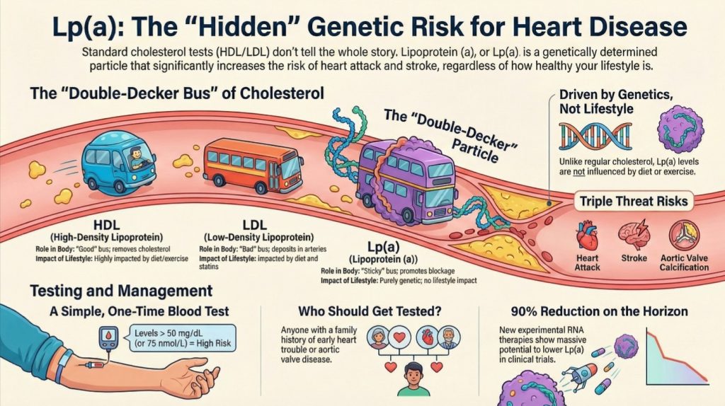
Geen enkele moderne discussie over lipide-gedreven cardiovasculaire aandoeningen is compleet zonder lipoproteïne(a), oftewel Lp(a). Structureel gezien is Lp(a) een LDL-achtig deeltje waaraan een extra apolipoproteïne(a) is gehecht. Klinisch gezien is het een van de belangrijkste erfelijk bepalaarde cardiovasculaire risicofactoren [1-3,5,9].
Ongeveer één op de vijf mensen heeft een verhoogd Lp(a), en de waarden zijn grotendeels genetisch bepaald. In tegenstelling tot standaard LDL wordt Lp(a) niet significant verbeterd door dieet of lichaamsbeweging en wordt het slechts in bescheiden mate beïnvloed door de meeste conventionele lipidenbehandelingen [1-3,5,9].
Lp(a) lijkt vooral gevaarlijk omdat het niet alleen atherogeen is, maar ook pro-inflammatoir en pro-trombotisch. Verhoogde spiegels zijn gekoppeld aan vroegtijdige myocardinfarct, beroerte, en aortaklep stenose. Om deze reden raden veel experts nu aan om bij iedere patiënt minstens één keer in het leven de Lp(a)-waarde te laten meten1-3,5,9].
Dit is een belangrijk bericht voor de volksgezondheid. Iemand kan veel dingen goed doen en toch een aanzienlijk erfelijk risico dragen door een verhoogd Lp(a). Dat risico vroegtijdig identificeren is van belang.
Waarom ApoB een beter klinisch kompas is dan LDL-C
Voor de dagelijkse praktijk is de belangrijkste diagnostische les eenvoudig: ApoB staat dichter bij de biologie van de ziekte dan LDL-C [1-3].
LDL-C schat hoeveel cholesterolmassa aanwezig is in LDL-deeltjes. ApoB schat hoeveel atherogene deeltjes er circuleren. Omdat elk deeltje één potentiële indringer is in de vaatwand, brengt ApoB de kans op arteriële beschadiging beter in kaart.
Niet-HDL-cholesterol is vaak een betere surrogaatmarker dan LDL-C, omdat het de cholesterol bevat die door alle ApoB-bevattende deeltjes wordt gedragen. Maar ApoB blijft de zuiverdere, op deeltjes gebaseerde meting. Een kransslagaderkalkscore kan helpen om latere manifestaties van gevestigde ziekte op te sporen, en Lp(a) helpt erfelijk risico te identificeren. Maar om de causale last van circulerende atherogene deeltjes te begrijpen, staat ApoB centraal [1-3].
Dit betekent niet dat LDL-C nutteloos is. Het blijft klinisch waardevol en algemeen beschikbaar. Maar wanneer het doel is om tests af te stemmen op het mechanisme, is ApoB de sterkere merker.
Het sluit ook beter aan bij de hierboven beschreven fysiologie. Als weefsels geen grote hoeveelheden circulerend cholesterol nodig hebben om te overleven, en als in de cholesterolbehoefte grotendeels wordt voorzien door lokale synthese en strikt gereguleerde intracellulaire routes, dan wordt de klinisch belangrijke vraag niet “Hoeveel cholesterol is er aanwezig in het plasma?”, maar “Hoeveel atherogene deeltjes wisselwerkingen voortdurend aan met de vaatwand?” ApoB beantwoordt die vraag beter dan LDL-C2,11-17].
Therapeutische implicaties: Vandaag en morgen
Huidige cholesterolverlagende therapie blijft enorm belangrijk. Statines, ezetimib, en PCSK9-remmers atherogene werking verminderen deeltjesbelasting en sterke resultaatgegevens hebben. PCSK9 remmers verlagen Lp(a) ook enigszins, en nieuwere op RNA gebaseerde therapieën die zich richten op Lp(a) laten in studies dramatische verlagingen zien [1,3,9].
Levensstijl blijft fundamenteel. Gewichtsverlies, verbeterd insulinegevoeligheid, een verminderde hepatische VLDL-productie en een lagere ontstekingsbelasting dragen allemaal bij aan het verminderen van het systemische risico [1]. Maar de opkomende biologie suggereert dat de toekomst van cardiovasculaire preventie mogelijk niet ophoudt bij het verlagen van de deeltjes in het plasma. Het kan ook inhouden dat de vaatwand direct wordt aangepakt.
Dat is waar de biologie van Rho-GEF bijzonder interessant wordt. Als specifieke GEF's zoals DOCK4 of SGEF de belangrijkste regulatoren zijn van LDL-opname of leukocytenrecruitment, dan zou directe remming van die routes een selectievere manier kunnen bieden om de vorming van plaque te verstoren4]. In plaats van alleen het verkeer op de weg te verminderen, zouden clinici op een dag ook de poort kunnen vernauwen en de ontstekingsreactie in de vaatwand kunnen afremmen.
Dit is nog een opkomend gebied, geen routinematige klinische strategie. Maar conceptueel wijst het in de richting van precisiegeneeskunde: het aanpakken van de specifieke lokale routes die circulerend risico omzetten in arteriële aandoeningen.
Het geïntegreerde raamwerk voor cholesterolhomeostase helpt ook te verklaren waarom agressieve verlaging van de ApoB-belasting biologisch plausibel is. Als aan de intracellulaire behoeften nog steeds kan worden voldaan via endogene synthese en streng gereguleerde weefselverwerking, dan hoeft het verminderen van circulerende atherogene deeltjes geen cellulaire onthouwing te betekenen. Het kan simpelweg de onnodige arteriële blootstelling verminderen2,11-17].
Een betere manier om aderverkalking uit te leggen
Voor het grote publiek kan de ziekte worden samengevat op een manier die zowel intuïtief als accuraat is.
ApoB-bevattende lipoproteïnen zijn te vergelijken met vrachtwagens die brandstof en structurele lading door de bloedbaan vervoeren. Als er te veel vrachtwagens op de weg zijn, belanden er op een gegeven momenten enkele in de wand van de slagaders. Zodra ze daar vast komen te zitten, raken ze beschadigd en kleverig. Het immuunsysteem stuurt opruimcellen af, maar die cellen raken overbelast en vormen plaque. Ondertussen is de slagaderwand zelf niet passief. Deze beschikt over moleculaire poortwachters die de toetreding van deeltjes, immuun-docking, cholesterolverwerking en weefselremodellering reguleren [1-8,11-15].
Dat raamwerk is eenvoudig genoeg voor het openbaar onderwijs, maar gedetailleerd genoeg om nuttig te zijn voor clinici. Het vermijdt de misleidende oversimplificatie dat cholesterol alleen slagaders “verstopt”, terwijl het de kernwaarde behoudt dat cholesterolbevattennde ApoB-deeltjes causaal noodzakelijk zijn voor de ziekte1-3].
Het laat ook ruimte voor een belangrijke nuance: het lichaam heeft cholesterol nodig, maar slechts in zeer kleine, streng gereguleerde hoeveelheden. Het echte gevaar is niet het bestaan van cholesterol zelf. Het gevaar is de chronische circulatie, binnendringing in de slagaders en retentie van ApoB-bevattende deeltjes in een weefselomgeving die hun lading omzet in ontsteking en plaque.
Conclusie
Atherosclerose kan het best worden begrepen als het snijvlak van twee systemen. Het eerste is de systemische lipoproteïnebiologie, waarbij ApoB-bevattende deeltjes circuleren als de noodzakelijke dragers van triglyceriden en cholesterol. Het tweede is de lokale arteriële biologie, waar endotheelcellen, leukocyten, macrofagen en gladde spiercellen bepalen of die deeltjes plaque worden.
ApoB weerspiegelt het aantal circulerende atherogene partikels en daarmee de statistische kans op arteriële beschadiging. Rho GEF–Rho GTPase-signaaltransductie helpt te verklaren waarom en hoe die beschadiging optreedt in de vaatwand. Samen bieden deze systemen een rijker ziektemodel dan het verouderde kader dat uitsluitend op LDL-C is gebaseerd.
Tegelijkertijd wordt cholesterol zelf gereguleerd door een streng gecontroleerde intracellulaire economie. Via het SREBP-2–SCAP–INSIG-systeem, de mevalonaatroute, LXR-gemedieerde effluxsignalisatie en weefselspecifieke synthese handhaaft het lichaam de kleine hoeveelheden cholesterol die het nodig heeft, terwijl het zichzelf verdedigt tegen een teveel [11-17]. Dit betekent dat de fysiologische behoefte aan cholesterol reëel is, maar de benodigde hoeveelheid is bescheiden, lokaal gereguleerd en fundamenteel anders dan de pathologische last die wordt opgelegd door overtollige circulerende ApoB-deeltjes.
Voor clinici ondersteunt dit een sterkere nadruk op ApoB, Lp(a) en vroege preventie. Voor het grote publiek maakt dit duidelijk dat hart- en vaatziekten niet eenvoudigweg gaan over het eten van vet of het hebben van “hoog cholesterol”. Het gaat over hoeveel atherogene deeltjes er circuleren, hoe lang ze aanwezig blijven, hoe de vaatwand daarop reageert, en hoe stoornissen in de lokale cholesterolverwerking achtergebleven deeltjes omzetten in ontsteking en plaque.
De toekomst van de lipidologie kan daarom tweeledig zijn: het verlagen van het aantal gevaarlijke deeltjes in de bloedbaan en tegelijkertijd het aanpakken van de moleculaire programma's die die deeltjes in de eerste plaats in de vaatwand laten binnendringen en beschadigen1-10In dat brede kader is een van de belangrijkste conceptuele correcties deze: het lichaam heeft geen groot circulerend overschot aan cholesterol nodig om te overlezen. Het heeft slechts een zeer kleine, zorgvuldig gereguleerde hoeveelheid op cellulair niveau nodig. Aderverkalking begint wanneer het transportsysteem die behoefte overschrijdt en de vaatwand daarvan de prijs betaalt.
Referenties
- Perone F, Bernardi M, Spadafora L, et al. Non-Traditional Cardiovascular Risk Factors: Tailored Assessment and Clinical Implications. J Cardiovasc Dev Dis. 2025;12(5):171. Published 2025 Apr 28. doi:10.3390/jcdd12050171
- Attia P, Dayspring T. #20 – Tom Dayspring, M.D., FACP, FNLA – Part I of V: an introduction to lipidology.
- Hill S. What Causes Cardiovascular Disease? Lipid Series Part 1. The Proof with Simon Hill.
- Li M, Jiao Q, Xin W, et al. The Emerging Role of Rho Guanine Nucleotide Exchange Factors in Cardiovascular Disorders: Insights Into Atherosclerosis: A Mini Review. Front Cardiovasc Med. 2022;8:782098. Published 2022 Jan 3. doi:10.3389/fcvm.2021.782098
- Heart Matters. Cholesterol Explained: HDL, LDL, and the Hidden Risk of Lp(a).
- Hackethal V. Research Suggests LDL Is Actively Transported Into Arterial Walls. NeurologyLive.
- Getz GS, Reardon CA. The “HDL receptor” scavenger receptor class B type 1 finesses the uptake of low-density lipoproteins into the subendothelial space of arteries. Biotarget.
- Huang L, Chambliss KL, Gao X, et al. SR-B1 drives endothelial cell LDL transcytosis via DOCK4 to promote atherosclerosis. Nature. 2019;569(7757):565-569. doi:10.1038/s41586-019-1140-4
- Nutrition Made Simple. Everything you need to know about Lp(a) | ft. Dr. Tom Dayspring.
- Heart Fit Clinic. Webinar on Cholesterol from a Lipidologist. Dr. Tom Dayspring.
- Brown MS, Goldstein JL. The SREBP pathway: regulation of cholesterol metabolism by proteolysis of a membrane-bound transcription factor. Cell. 1997;89(3):331-340. doi:10.1016/s0092-8674(00)80213-5
- Brown MS, Goldstein JL. Sterol regulatory element binding proteins (SREBPs): controllers of lipid synthesis and cellular uptake. Nutr Rev. 1998;56(2 Pt 2):S1-S75. doi:10.1111/j.1753-4887.1998.tb01680.x
- Muneer PMA, Alikunju S, Mishra V, et al. Activation of NLRP3 inflammasome by cholesterol crystals in alcohol consumption induces atherosclerotic lesions. Brain Behav Immun. 2017;62:291-305. doi:10.1016/j.bbi.2017.02.014
- Feng B, Yao PM, Li Y, et al. The endoplasmic reticulum is the site of cholesterol-induced cytotoxicity in macrophages. Nat Cell Biol. 2003;5(9):781-792. doi:10.1038/ncb1035
- Murphy AJ, Akhtari M, Tolani S, et al. ApoE regulates hematopoietic stem cell proliferation, monocytosis, and monocyte accumulation in atherosclerotic lesions in mice. J Clin Invest. 2011;121(10):4138-4149. doi:10.1172/JCI57559
- Dietschy JM, Turley SD. Cholesterol metabolism in the brain. Curr Opin Lipidol. 2001;12(2):105-112. doi:10.1097/00041433-200104000-00003
- Miller WL. MECHANISMS IN ENDOCRINOLOGY: Rare defects in adrenal steroidogenesis. Eur J Endocrinol. 2018;179(3):R125-R141. doi:10.1530/EJE-18-0279
- Ginsberg HN. Lipoprotein metabolism and its relationship to atherosclerosis. Med Clin North Am. 1994;78(1):1-20. doi:10.1016/s0025-7125(16)30174-2

