Structure, transport et rôle biologique du cholestérol
Une analyse mécanistique de la répartition entre compartiments, lipoprotéine les exigences structurelles, ainsi que la valeur minimale estimée par le modèle pour le système de transport en circulation
Résumé
Contexte. Tout le corps cholestérol La distribution du cholestérol, les besoins structurels des lipoprotéines et la dépendance des tissus vis-à-vis du cholestérol circulant sont souvent confondus dans les sources cliniques et pédagogiques, ce qui conduit à surestimer la quantité de cholestérol circulant dont l'organisme “ a besoin ”.”
Méthodes. Cette analyse (i) définit un vocabulaire rigoureux, basé sur les compartiments, qui distingue le cholestérol total de l'organisme, le cholestérol tissulaire, le cholestérol intracellulaire et le cholestérol circulant (plasma/sérum), et (ii) élabore une estimation transparente, fondée sur un modèle de transport, de la concentration minimale en cholestérol circulant nécessaire pour maintenir l’intégrité structurelle des lipoprotéines et répondre à la demande tissulaire exogène, avec des hypothèses clairement énoncées, des paramètres explicitement convertis en unités et des limites de sensibilité. Les équations et les plages de paramètres sont présentées dans l’annexe « Méthodes » (section 11).
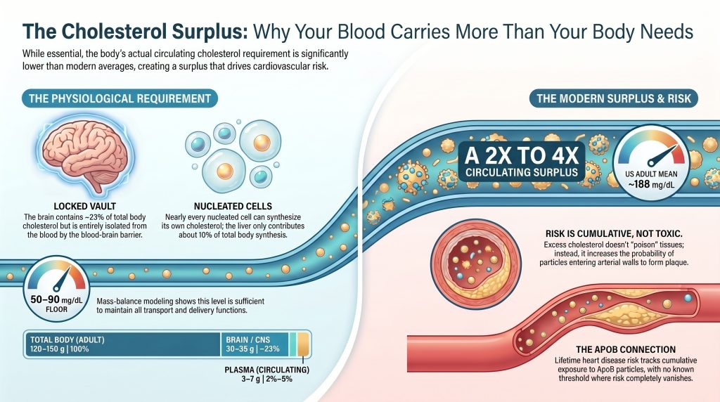
Résultats. (1) La quantité totale de cholestérol dans l'organisme d'un adulte est d'environ 120 à 150 g ; le pool plasmatique à renouvellement rapide représente 3 à 7 g (en fonction du volume plasmatique et de la mesure cholestérol total (concentration) représente 2 à 51 TP9T de la masse corporelle. (2) Le cerveau à lui seul en contient environ 30 à 35 g, isolés du plasma par la barrière hémato-encéphalique. (3) Le cholestérol plasmatique se présente sous forme d’environ 70% de cholestérol estérifié et d’environ 30% de cholestérol libre. (4) Une modélisation représentative à 22 nm LDL Une particule (reconstruction de Hevonoja) contient environ 2 200 molécules de cholestérol, dont environ 181 TP9T correspondent à du cholestérol libre en surface ; les LDL sont hétérogènes et ces chiffres ne doivent pas être appliqués sans discernement à toutes les particules de LDL. (5) Selon les hypothèses énoncées dans le modèle de transport et en utilisant des comptages de particules compatibles avec la RMN (LDL-P en nmol/L → environ 10¹⁶–10¹⁷ particules/dL ; HDL-P en µmol/L → environ 10¹⁸ particules/dL), le taux plasmatique minimal de cholestérol total estimé par le modèle, qui stabilise simultanément le système lipoprotéique et couvre environ 50 mg/jour de la demande exogène nette chez un adulte non enceinte, est d’environ 50–90 mg/dL (plage de sensibilité : 40–110 mg/dL). Il s’agit d’un scénario théorique, et non d’un minimum physiologique avéré.
Conclusions. Le taux moyen actuel de cholestérol total plasmatique chez les adultes américains, qui s’élève à environ 188 mg/dL (NHANES 2017-2018), dépasse le seuil minimal de transport estimé par le modèle d’un facteur qui dépend des hypothèses retenues, mais qui est systématiquement supérieur à 2. Cet excédent est toléré sur le plan métabolique, il est causalement lié au risque d’ASCVD sur la base de chaque particule d’apoB, et il n’est nécessaire à aucune fonction physiologique connue. Les recommandations cliniques préconisant une réduction du LDL-C pour la prévention de l’ASCVD s’inscrivent dans un registre épistémique différent (réduction des événements dans les essais cliniques) de celui de cet article (modélisation du bilan massique) ; ces deux éléments sont complémentaires, et non contradictoires.
1. Vocabulaire par compartiment
Chaque affirmation relative au cholestérol figurant dans cet article précise (i) le compartiment, (ii) la forme chimique et (iii) les unités. La confusion entre les concentrations cliniques des lipides et la masse de cholestérol tissulaire ou corporelle est à l'origine de la plupart des erreurs quantitatives présentes dans la littérature grand public et pédagogique. La terminologie suivante est utilisée tout au long de cet article. [1]
| Terme | Signification | Unités courantes |
| Colestérol plasmatique / colestérol sérique | Taux de cholestérol total dans la fraction sanguine correspondante (calculé ou mesuré directement dans le cadre d'un bilan lipidique de routine) | mg/dL ou mmol/L |
| LDL-C / HDL-C / VLDL-C plasmatiques | Cholestérol transporté par la classe de lipoprotéines mentionnée dans le plasma | mg/dL ou mmol/L |
| LDL-P / HDL-P | Concentration en particules de LDL ou HDL particules (généralement mesurées par spectroscopie RMN) | LDL-P en nmol/L ; HDL-P en µmol/L (à noter la différence d'ordre de grandeur) |
| Cholestérol libre plasmatique / plasma ester de cholestérol | Répartition chimique du cholestérol plasmatique entre les formes non estérifiées et estérifiées (généralement ~30% / ~70%) | mg/dL, mmol/L ou % de cholestérol total plasmatique |
| Masse de cholestérol plasmatique | TC plasmatique × volume plasmatique ; masse absolue de cholestérol circulant dans le plasma à un instant donné | g (= mg/dL × dL de plasma / 1 000) |
| Masse de cholestérol circulant | Masse de cholestérol plasmatique + cholestérol présent dans les membranes des érythrocytes (qui s'échange rapidement avec le plasma) | g |
| Concentration de cholestérol dans les tissus | Taux de cholestérol par unité de masse tissulaire (par exemple, dans le cerveau : environ 23 mg/g de poids humide) | mg/g de poids humide ou sec |
| Cholestérol cellulaire / subcellulaire | Le cholestérol présent dans un type de cellule, une membrane d'organite ou un réservoir spécifique | mol% de lipides membranaires ; µg/mg protéine |
| Cholestérol total | Masse totale de cholestérol dans l'ensemble des tissus et des liquides de l'organisme | g |
| Seuil de transport estimé par le modèle | Concentration plasmatique minimale de cholestérol calculée permettant d'assurer les fonctions structurelles et de transport du système des lipoprotéines circulantes, selon les hypothèses énoncées | mg/dL |
| ■ Les unités de particules de lipoprotéines sont des conventions utilisées en RMN
L'analyse des sous-fractions lipoprotéiques par RMN (LipoProfile et autres méthodes similaires) exprime la concentration en particules de LDL (LDL-P) en nanomoles (nmol/L), ce qui reflète la fourchette typique chez l'adulte, comprise entre environ 10¹⁵ et 10¹⁸ particules/L. La concentration en particules de HDL (HDL-P) est généralement exprimée en micromoles (µmol/L), car les particules de HDL sont plus abondantes. Cet article utilise les conventions de la RMN tout au long du texte et effectue la conversion en particules par dL à l'aide de calculs explicites. |
| ■ Plasma ou sérum ?
Les bilans lipidiques cliniques de routine sont réalisés soit sur du sérum, soit sur du plasma ; la différence entre les valeurs de cholestérol mesurées est faible (généralement < 3%). Sauf indication contraire, toutes les concentrations de cholestérol circulant font référence indifféremment au plasma ou au sérum. |
2. Répartition systémique et bilan massique
2.1 Cholestérol total
Le taux de cholestérol total chez un adulte est d'environ 120–150 g, avec mise à l'échelle en fonction du poids corporel. Cette fourchette reflète la variabilité inter-sujets et inter-méthodes observée dans les études sur la répartition tissulaire. [2][3] Les références tirées d’anciens manuels, qui mentionnent une valeur d’environ 35 g, ne concernent que le « Pool A » à échange rapide de Goodman (foie + plasma + érythrocytes + une partie des viscères) dans le modèle cinétique des traceurs, et non la teneur totale de l’organisme. Des analyses tissulaires quantitatives directes (Sabine, 1977, reprises dans des revues récentes) situent le cholestérol total du corps chez l’adulte à un niveau plus élevé, le cerveau, les tissus conjonctifs (y compris les tissus adipeux) et les muscles squelettiques y contribuant chacun à hauteur d’environ 25 à 35 g. [2][3]
Le modèle de Goodman à trois réservoirs définit un réservoir A à échange rapide (foie, plasma, érythrocytes, tissus splanchniques) de 15 à 30 g, un réservoir B à échange lent (muscles squelettiques, tissu adipeux, tissu conjonctif dense) de 35 à 60 g, et un pool du SNC cinétiquement isolé d’environ 30 à 35 g. [4][5] Le compartiment plasmatique à lui seul contient 3 à 7 g de cholestérol, en fonction du volume plasmatique et de la concentration plasmatique mesurée de cholestérol total.
2.2 Répartition tissulaire
Répartition calculée en partant d'une hypothèse de ~140 g de cholestérol total dans l'organisme (valeur médiane représentative). Toutes les données correspondent à la masse de cholestérol tissulaire. [2][3]
| Compartiment | Cholestérol (g) | % du poids total du corps | Forme chimique dominante |
| Cerveau / SNC (myéline + membranes neuronales) | 30–35 | ~22–25% | >99,51 TP9T libre (non estérifié) |
| Muscle squelettique | ~25–30 | ~18–22% | Libre (membrane plasmique) |
| Tissu conjonctif, tissu adipeux, liquides organiques | ~25–32 | ~18–23% | Mixte ; l'accumulation de CE augmente avec l'âge dans les tendons et la dure-mère |
| Peau | ~12–18 | ~9–13% | Gratuit |
| Autres viscères (poumons, reins, intestins, etc.) | ~12–18 | ~9–13% | Principalement gratuit |
| Foie | ~4–6 | ~3–4% | FC + CE mixtes |
| Plasma / pool circulant | 3–7 | ~2–5% | ~70% CE / ~30% FC |
| Cœur, rate, glandes surrénales (par organe) | <1 de chaque | <1% | Haute densité par gramme (glandes surrénales ≥ 25 mg/g) |
Le SNC contient environ 22 à 251 TP9T de cholestérol corporel, avec une concentration tissulaire maximale d’environ 23 mg/g de poids humide. [5][6] La barrière hémato-encéphalique est imperméable au cholestérol lié aux lipoprotéines; l'ensemble du pool de cholestérol du SNC est synthétisé localement. Le cholestérol des neurones corticaux est renouvelé avec une demi-vie comprise entre 6 mois et 5 ans ; celui de la myéline est renouvelé à l'échelle de plusieurs décennies. L'efflux net hors du SNC se produit principalement sous forme de 24S-hydroxycholestérol, qui traverse la barrière hémato-encéphalique et est éliminé dans le plasma. [5][6][7]
2.3 Masse de cholestérol plasmatique à différentes concentrations de cholestérol total plasmatique
La masse de cholestérol plasmatique correspond au produit du volume plasmatique par la concentration plasmatique en cholestérol total. Pour un volume plasmatique représentatif de 3,0 L :
| TC plasmatique (mg/dL) | Masse de cholestérol plasmatique (g, 3,0 L de plasma) | Contexte clinique |
| 50 | 1.5 | En dessous de la fourchette néonatale normale ; mutations LOF rares |
| 80 | 2.4 | Signalé PCSK9 Plage des homozygotes LOF |
| 100 | 3.0 | Inhibiteur de PCSK9 objectifs pendant le traitement |
| 150 | 4.5 | Objectif thérapeutique en matière de traitement hypolipidémiant intensif |
| 188 | 5.6 | Moyenne chez les adultes américains (NHANES 2017-2018) [8] |
| 220 | 6.6 | Légèrement supérieur à la normale |
| 280 | 8.4 | Intervalle de valeurs pour la FH hétérozygote |
| ■ Le volume plasmatique varie en fonction de la taille corporelle
Le volume plasmatique chez l'adulte est d'environ 40 à 45 ml/kg de poids corporel (environ 2,8 à 3,2 L pour un adulte de 70 kg, environ 3,5 à 4,0 L pour un adulte de 90 kg). Sauf indication contraire, tous les calculs de la masse plasmatique présentés dans cet article utilisent une valeur représentative de 3,0 L. |
2.4 Distribution subcellulaire et capteur de cholestérol du réticulum endoplasmique
Dans la plupart des cellules non neuronales, la membrane plasmique contient entre 60 et 90% de cholestérol cellulaire pour 30 à 40 mol% de lipides membranaires, tandis que le réticulum endoplasmique n'en contient que 3 à 6 mol%. [9][10] Ce gradient est régulé par la protéine SREBP2, associée à SCAP et à Insig : lorsque la concentration en cholestérol dans le réticulum endoplasmique (RE) dépasse environ 5 mol%, SCAP retient SREBP2 dans le RE, ce qui entraîne une inhibition de la synthèse. [11] Das et al. (eLife 2014) ont identifié trois pools fonctionnels de cholestérol dans la membrane plasmique à l’aide d’une sonde à la Perfringolysine O (PFO*) : un pool séquestré dans la sphingomyéline (~15 mol% de lipides de la membrane plasmique), un pool “ essentiel ” nécessaire à la viabilité cellulaire, et un pool “ accessible ” qui circule vers le réticulum endoplasmique pour réguler la synthèse. [11][12] C'est la quantité de cholestérol disponible — et non le cholestérol PM en vrac — qui constitue le signal régulateur.
| ▲ Le seuil de liaison du PFO* s'applique aux bicouches de membrane plasmique, et non aux monocouches de lipoprotéines.
Le seuil d'accessibilité de la mol% PFO* à 35 dans les membranes plasmiques reflète l'architecture de la bicouche et la complexation sphingomyéline-cholestérol spécifique à la membrane plasmique. [11][12] Les surfaces des LDL et des HDL sont constituées de monocouches de phospholipides stabilisées par apolipoprotéines; elles présentent une composition lipidique différente, une asymétrie des feuillets et des contraintes d'empilement. Ce seuil ne doit pas être appliqué directement aux surfaces des lipoprotéines sans analyse complémentaire. |
2.5 Chiffre d'affaires quotidien
De novo pour l'ensemble du corps synthèse du cholestérol chez les adultes, elle est d'environ 10 mg/kg/jour (environ 700 mg/jour chez un adulte de 70 kg, environ 880 mg/jour chez un adulte de 88 kg). Le foie ne participe qu'à hauteur d'environ 10% à la synthèse totale chez l'être humain ; la majeure partie de cette synthèse est extra-hépatique. [13] Des études de traceurs à trois réservoirs portant sur le renouvellement du cholestérol plasmatique indiquent un taux de production total (synthèse et absorption alimentaire) d'environ1,0 à 1,2 g/jour chez les sujets normolipidémiques. [4] L'apport alimentaire (environ 300 à 500 mg/jour dans le cadre d'un régime occidental, dont environ 30 à 50% sont absorbés) est compensé par une régulation à la baisse de la synthèse endogène. [14]
3. Architecture des lipoprotéines et répartition du cholestérol
3.1 Composition d'une particule représentative de LDL de 22 nm
La reconstitution ci-dessous est tirée de Hevonoja et al. (BBA 2000) et concerne une particule LDL représentative d'un diamètre de 22 nm. [15][16][17]
| Composant | Molécules / particules | Fraction massique (%) | Lieu |
| Esters de cholestéryle (CE) | ~1,600 | 40–45% | Noyau |
| Cholestérol non estérifié (FC) | ~600 | 8–10% | environ 400 en surface, environ 200 en profondeur |
| Triglycérides (TG) | ~170 | 5–9% | Noyau |
| Phosphatidylcholine | ~450 | } 19–21% combiné | Monocouche de surface |
| Sphingomyéline | ~185 | (phospholipides totaux) | Monocouche de surface |
| ApoB-100 (un seul exemplaire) | 1 | 20–25% | Enveloppe la surface |
Le cholestérol total par particule LDL représentative est d'environ 2 200 molécules ; le FC de surface représente environ 18% de cholestérol LDL. Certaines sources générales mentionnent environ 1 500 de cholestérol par particule moyenne de LDL, ce qui reflète les différentes méthodes de calcul de la moyenne utilisées pour la distribution des tailles des LDL ; ces deux chiffres constituent des descriptions de la tendance centrale d'une population intrinsèquement hétérogène. [16]
| ▲ Le LDL est hétérogène.
In vivo, la taille des LDL varie entre environ 22 et 27,5 nm, avec des variations importantes de leur composition ; petites densités de LDL contient moins de cholestérol par particule et est riche en triglycérides, tandis que les grosses particules de LDL flottantes en contiennent davantage. La reconstruction de Hevonoja correspond à une particule représentative de la tendance centrale et ne doit pas être appliquée sans discernement à toutes les espèces de LDL. |
3.2 Rapport entre la fraction libre et la fraction estérifiée dans le plasma
Des mesures enzymatiques et chromatographiques directes réalisées sur du sérum humain sain montrent que le cholestérol libre dans le plasma représente environ 25 à 30% du cholestérol plasmatique total, la fraction estérifiée représentant quant à elle environ 70 à 75%. [18][19] Un rapport FC/CE élevé est associé à un dysfonctionnement de la LCAT, à une chylomicronémie familiale et, selon certaines études, à un facteur athérogène indépendant. [20]
3.3 Cholestérol libre de surface : rôle structurel
Le cholestérol libre à la surface des lipoprotéines s'intercale entre les chaînes acyles des phospholipides, son groupe 3β-hydroxyle faisant saillie dans l'interface aqueuse et sa structure rigide stérol anneau aligné avec les queues lipidiques. Cette “ condensation ” réduit le volume libre dans la monocouche, diminue sa perméabilité à l'eau et augmente la stabilité mécanique de la particule. [21][22] Une concentration en FC d'environ 25 mol% de lipides de surface est compatible avec une monocouche ordonnée et bien compacte ; cette configuration favorise la stabilité des particules et leur mobilité latérale, ce qui est nécessaire au traitement enzymatique. [23]
3.4 Échange de cholestérol libre
Le cholestérol libre à la surface des lipoprotéines est en équilibre spontané avec les membranes cellulaires et d’autres particules lipoprotéiques par diffusion passive dans le milieu aqueux. Le temps de demi-vie d’échange est inversement proportionnel à la courbure de la surface : environ 5 minutes pour les HDL naissantes, environ 45 minutes pour les LDL. [21][23] Ce système cinétique assure une activité thermodynamique quasi uniforme du cholestérol sur l'ensemble des surfaces lipidiques extracellulaires. [22][23]
4. Esters de cholestérol : le transport des substances
Les esters de cholestéryle sont entièrement hydrophobes et se trouvent au cœur des lipoprotéines sous forme de gouttelettes liquides ou à l'état de cristaux liquides. Les esters de cholestéryle (CE) présents dans les LDL représentent 40 à 45% de la masse des particules et constituent la majeure partie du cholestérol acheminé vers les cellules par endocytose médiée par des récepteurs, où la lipase acide lysosomale libère le cholestérol libre en vue de son utilisation. [17]
CETP transporte la CE de l'HDL vers les lipoprotéines contenant de l'apoB en échange de triglycérides, ce qui enrichit apoB particules transportant du cholestérol. Une part importante du CE du noyau des particules d’apoB correspond donc au cholestérol ayant circulé dans les cellules périphériques, le plasma et les HDL avant d’être transféré vers les LDL, s’ajoutant ainsi aux réserves hépatiques de cholestérol sécrétées sous forme de VLDL. [24]
5. Maturation des HDL
Le HDL naissant est sécrété sous forme d'apoA-I pauvre en lipides, qui acquiert des phospholipides et du cholestérol libre à partir des cellules périphériques par le biais de ABCA1, formant une particule discoïdale composée de deux molécules d'apoA-I dans une conformation en “ double ceinture ”. [25] La LCAT, activée par l'apoA-I, transfère un groupe acyle de la position sn-2 de la phosphatidylcholine vers la FC de surface, générant ainsi de la CE qui migre vers le cœur hydrophobe. [23][25]
À mesure que le CE s'accumule, la bicouche discoïdale prend une forme sphérique. Une particule de HDL2 mature atteint un diamètre d'environ 10 à 12 nm et contient environ 30 à 60 molécules de cholestérol par particule (principalement du CE), avec environ 5 à 15 molécules de FC en surface. [23] La CE retourne soit directement vers le foie par le biais de SR-B1 ou est transféré vers des particules d’apoB par l’intermédiaire de la CETP en vue de sa clairance hépatique par le biais de LDLR. [23][24]
6. Besoins tissulaires : synthèse de novo et absorption exogène
6.1 La plupart des tissus nucléés sont capables de synthétiser le cholestérol de novo
Chaque cellule nucléée du corps humain exprime l'ensemble de la voie de biosynthèse du mégalonate et du cholestérol. [13][26] Dans des conditions normales, la plupart des tissus ne dépendent pas totalement d'un apport continu en cholestérol exogène provenant du plasma ; l'équilibre quantitatif entre la synthèse locale et l'absorption dérivée des lipoprotéines varie selon le tissu, l'âge et l'état physiologique.
| Tissu | Source principale de cholestérol | Remarques |
| Cerveau / Système nerveux central | Synthèse locale (astrocytes → les neurones via apoE) | Le BBB empêche le passage du cholestérol lié aux lipoprotéines ; efflux net via le 24S-OH-cholestérol [7] |
| Muscle squelettique | Synthèse essentiellement locale | Faible demande pour ce nouveau stérol |
| Peau | ~90% : synthèse locale | Favorise la fonction barrière |
| Cortex surrénal | Résultats mitigés : LDLR > SR-B1 chez l'homme ; l'inverse chez les rongeurs [27][28] | Les réserves de CE répondent à une demande stéroïdogénique aiguë |
| Gonades (testicules, ovaires) | Mixte : LDLR + SR-B1 ; dépendant du statut reproductif | La grossesse et l'allaitement augmentent les besoins |
| Placenta | LDL et HDL maternels ; grossesse uniquement | Hors du champ d'application de l'analyse portant sur les adultes non enceintes |
| Foie | Synthèse + apport alimentaire + transport inverse | Régulateur principal ; sécrète des VLDL |
Dans la physiologie adrénocorticale et gonadique humaine, l’endocytose médiée par le récepteur LDL (LDLR) joue un rôle quantitatif plus important que le SR-B1 dans l’apport en cholestérol — contrairement au modèle observé chez les rongeurs, où le SR-B1 domine. Le SR-B1 reste néanmoins exprimé et biologiquement actif dans les tissus stéroïdogènes humains, et une perte de fonction du SR-B1 chez l'homme entraîne des anomalies subtiles mais réelles de la réponse du cortisol stimulée par l'ACTH. La nature mixte des voies d'apport du cholestérol aux tissus stéroïdogènes humains doit être prise en compte dans toute analyse mécanistique. [27][28]
6.2 Cinétique des récepteurs LDL : les études in vitro et in vivo ne sont pas interchangeables
In vitro : Dans les fibroblastes humains en culture, la liaison à haute affinité de l'apoB-100 des LDL atteint sa saturation à des concentrations en protéines LDL inférieures à 50 µg/mL (Brown & Goldstein). [29][30] Catapano et ses collaborateurs (2024) décrivent ce phénomène comme une demi-saturation correspondant à un équivalent plasmatique de LDL-C d’environ 2,5 mg/dL, et associent cette donnée à l’observation selon laquelle le LDL-C du liquide interstitiel représente environ 20% du LDL-C plasmatique pour affirmer qu’un taux de LDL-C plasmatique d’environ 12,5 mg/dL saturerait les LDLR tissulaires. [31] Cet argument repose sur des hypothèses intermédiaires concernant le rapport entre la masse protéique et la masse cholestérique des LDL, la composition du liquide interstitiel et l'uniformité du comportement des récepteurs LDL (LDLR) des fibroblastes ; cet article cite cet argument sans pour autant le valider comme seuil mesuré chez l'être humain.
In vivo : La clairance du LDL au niveau des organes chez le rat et le hamster (Spady & Dietschy) donne une valeur de K_m ≈ 90 mg/dL — soit environ 30 fois plus élevée, ce qui s'explique davantage par la présence de couches limites non agitées, la perméabilité capillaire et la densité des récepteurs que par l'affinité intrinsèque. [32] Cette valeur observée in vivo chez les rongeurs ne peut pas être directement transposée aux seuils d’apport en cholestérol au niveau des tissus humains.
| ▲ La cinétique de saturation des récepteurs ne constitue pas un seuil de distribution clair chez l'être humain.
Cette chaîne (Km des fibroblastes in vitro → LDL-C équivalent plasmatique → saturation en LDLR du liquide interstitiel dans les cellules surrénales/gonadiques) repose sur des hypothèses qui n’ont pas été validées de manière indépendante chez l’homme ; de plus, les hépatocytes sont exposés à du sang sinusoïdal plutôt qu’à du liquide interstitiel ordinaire. L'affirmation réellement défendable est d'ordre empirique : les personnes présentant un taux plasmatique de LDL-C très faible, par génétique ou ayant suivi un traitement, n'ont pas présenté de signes cliniques d'insuffisance d'apport en cholestérol dans les populations étudiées. |
6.3 Observations empiriques : taux de LDL-C très bas chez l'être humain
Les porteurs de mutations homozygotes de perte de fonction du gène PCSK9 constituent un petit échantillon rapporté. Les homozygotes afro-américains de l'étude « Dallas Heart Study », décrits à l'origine, présentaient tout au long de leur vie un taux plasmatique de LDL-C de 14 à 29 mg/dL (cholestérol total généralement compris entre 50 et 80 mg/dL) sans anomalies cliniques manifestes, d'après les rares descriptions publiées. [33][34] Le nombre total de ces homozygotes recensés à l'échelle mondiale reste faible ; ces données viennent donc étayer l'absence de risque majeur, mais ne prouvent pas l'existence d'un seuil physiologique minimal universel.
Les données les plus solides proviennent d'essais randomisés portant sur les inhibiteurs de la PCSK9. FOURIER (n = 27 564) ont présenté une durée médiane de suivi après randomisation de 2,2 ans, avec un taux médian de LDL-C sous traitement de 30 mg/dL et aucun signe d'insuffisance surrénale, d'hypogonadisme ou de déclin cognitif, accident vasculaire cérébral hémorragique, ou une maladie musculaire. [35] ODYSSEY Les résultats (n = 18 924) ont fait l'objet d'un suivi médian de 2,8 ans et ont montré des résultats similaires en matière de sécurité. [36] L'étude de prolongation en ouvert FOURIER (FOURIER-OLE, n = 6 635 sur les 27 564 initiaux) a ajouté une médiane 5,0 ans de évocumab exposition, entraînant exposition cumulative dans ce sous-groupe à environ 8,4 ans, sans qu'aucun nouveau signal de sécurité n'ait été observé. [37] Il s'agit là de données prospectives à grande échelle sur la sécurité ; elles ne constituent pas une mesure directe des seuils de tolérance du cholestérol chez l'être humain, mais elles délimitent par le haut la fourchette de sécurité empiriquement démontrée pour le LDL-C plasmatique.
Selon des études menées sur les lymphocytes périphériques humains, la concentration en LDL dans le liquide interstitiel serait comprise entre environ 10 et 201 TP9T de la concentration plasmatique en LDL. [38][39] Ces données sont de nature descriptive et ne permettent pas d'établir un seuil mesuré de saturation des tissus humains.
7. Estimation du taux minimal de cholestérol circulant à l'aide d'un modèle de transport
| ■ Ce que cette section est et ce qu'elle n'est pas.
Cette section présente une estimation, fondée sur un modèle de transport, de la concentration plasmatique minimale en cholestérol qui permet à la fois (i) de maintenir l'intégrité structurelle du système des lipoprotéines circulantes et (ii) de couvrir les besoins quotidiens nets en cholestérol exogène d'un adulte non enceinte dans des conditions normales. Il s'agit d'une estimation calculée et non d'une mesure. Les équations et les valeurs des paramètres figurent à la section 11. |
7.1 Intégrité structurelle
Toutes les particules de lipoprotéines circulantes ont besoin de cholestérol libre en surface pour stabiliser leur monocouche phospholipidique. À partir de comptages de particules compatibles avec la RMN (LDL-P en nmol/L ; HDL-P en µmol/L) et d’une conversion d’unités explicite : [15][17]
| Classe de particules | Spécialisation (clinique) | Particules par dL | Surface FC par particule | Contribution du FC de surface (mg/dL) |
| LDL | LDL-C : 100 mg/dL ; LDL-P : environ 1 200 nmol/L | ~7,1 × 10¹⁶ | ~400 | ~18 |
| HDL | HDL-C 50 mg/dL ; HDL-P ~30 µmol/L | ~1,8 × 10¹⁸ | ~10 | ~12 |
| VLDL / IDL | D'origine TG ; nombre de particules : environ 10¹⁵–10¹⁶ /dL | — | ~variable | ~2–3 |
| Surface totale de la FC (modèle, TC de référence ~190) | — | — | — | ~30–33 |
| FC plasmatique totale mesurée (~28% de TC à TC 190) | — | — | — | ~50–55 (comprend les FC principaux et autres) |
La concentration de FC en surface, estimée par le modèle, d’environ 30 à 33 mg/dL pour des valeurs lipidiques cliniques typiques, à laquelle s’ajoute la concentration de FC du noyau d’environ 10 à 15 mg/dL (le LDL présente une FC de base d’environ 200 par particule, tandis que le HDL apporte une contribution moindre en termes de FC de base), ainsi qu’une faible contribution des résidus de chylomicrons et de la Lp(a), donne un total d’environ 45 à 55 mg/dL de FC plasmatique. Ce résultat correspond à la fraction FC plasmatique mesurée directement, comprise entre environ 25 et 301 TP9T du cholestérol total, pour un taux de cholestérol total d'environ 190 mg/dL. [18][19]
7.2 Apport quotidien net de cholestérol d'origine exogène vers les tissus périphériques
Transport inverse du cholestérol Des études de traçage menées chez des adultes en bonne santé (Turner et al., 2012) estiment l'efflux de FC des tissus de l'ensemble du corps à 3,79 ± 0,88 mg/kg/h, ce qui a été rapporté dans l'article original comme étant d'environ 8 g/jour ; ce qui correspond à environ 6,4 g/jour pour un poids de 70 kg ou à environ 8 g/jour pour un poids de 88 kg (poids corporel moyen approximatif de l’étude). [40] Il s'agit ici d'un flux d'échange bidirectionnel, et non de la demande nette d'apport. L'absorption exogène nette estimée, indispensable aux tissus dont la synthèse locale est insuffisante dans des conditions de référence, chez une adulte non enceinte, est d'environ :
| Tissu / fonction | Consommation quotidienne estimée de cholestérol | Source d'approvisionnement |
| Stéroïdogenèse surrénale (valeur de référence) | environ 30 à 60 mg/jour | Principalement par l'intermédiaire du LDLR ; contribution du SR-B1 |
| Stéroïdogenèse gonadique (adulte non enceinte, valeur de référence) | < 10 mg/jour | Chez l'être humain, ce processus est principalement médié par le récepteur LDLR (LDLR) |
| Acide biliaire synthèse | environ 400 mg/jour | Réserve hépatique ; il ne s'agit pas d'une demande périphérique |
| Renouvellement des membranes extra-hépatiques (toutes) | environ 600 à 700 mg/jour | Obtenu par synthèse locale |
| Demande exogène nette obligatoire (adulte non enceinte, valeur de référence) | De l'ordre de quelques dizaines de mg/jour (estimation centrale d'environ 50 mg/jour) | Niveau très bas de LDL-C plasmatique |
| ▲ Adulte non enceinte, données initiales uniquement.
La grossesse entraîne environ un doublement du taux de cholestérol plasmatique chez la mère ; les besoins du placenta, d'un ordre de grandeur différent, ne sont pas pris en compte ici. Une maladie aiguë, un stress intense et une régénération tissulaire rapide augmentent également ces besoins. |
7.3 Seuil de transport estimé par le modèle combiné
Résultats du modèle pour une gamme de valeurs de TC du plasma :
| Scénario « Plasma TC » | FC de surface du LDL (mg/dL) | FC de la HDL (mg/dL) | FC plasmatique totale (mg/dL) | Remarques |
| TC 188 (moyenne américaine) | ~18 | ~12 | environ 50 à 55 | Ligne de base |
| TC 100 (cible PCSK9-Rx) | ~10 | ~10 | ~30 | Suffisant ; bien au-dessus de tout seuil structurel |
| TC 70 (homozygote pour une perte de fonction du gène PCSK9) | ~4 | ~9 | ~17 | Au niveau du plancher de référence ou à proximité ; toléré d'après l'expérience |
| TC 50 (génétique extrême) | ~2 | ~8 | ~13 | En dehors de tout cas empirique dûment validé |
En résumé : Le modèle de transport prévoit qu’un adulte non enceinte, dans des conditions de référence, peut maintenir un système fonctionnel de lipoprotéines circulantes et répondre à une demande tissulaire exogène d’environ 50 mg/jour pour un taux de cholestérol total plasmatique de l’ordre de 50 à 90 mg/dL (plage de sensibilité : 40–110 mg/dL). La forme chimique reste, dans cette plage, composée d’environ 70% estérifiée et 30% non estérifiée. L’estimation minimale du modèle concorde numériquement avec le taux de cholestérol total (CT) plasmatique observé chez les homozygotes porteurs d’une perte de fonction (LOF) du gène PCSK9 (~50–80 mg/dL), ce qui apporte une confirmation indépendante des observations cliniques et génétiques. Le taux moyen de CT plasmatique chez les adultes américains est actuellement d’environ 188 mg/dL [8] dépasse le seuil estimé par le modèle d'environ 2 à 4 fois, selon le scénario de sensibilité retenu.
7.4 Limites de sensibilité
| Hypothèses variables | Portée testée | Incidence sur l'estimation des coûts de transport |
| Volume plasmatique | 2,5 à 4,0 L | Échelle inverse ; effet principal sur la masse absolue, effet secondaire sur les mg/dL |
| LDL-P pour un taux donné de LDL-C | ± 50% de référence | Affecte le composant FC de manière proportionnelle |
| FC de surface par particule de LDL | 300–500 | ± 15% sur le composant FC |
| FC par particule de HDL | 5–15 | ± 25% sur le composant FC des HDL |
| Demande nette de biens périphériques | 20 à 150 mg/jour | Ajoute une marge de 5 à 30 mg/dL |
| Grossesse / maladie aiguë | — | Modèle non applicable ; la demande peut être multipliée par 2 à 10 |
| Plage plausible combinée (adulte non enceinte, valeur de référence) | — | ~40 à 110 mg/dL de cholestérol total (CT) plasmatique |
8. Causalité liée à l’ApoB et risque d’ASCVD
La question de savoir si les tissus périphériques ont besoin de cholestérol circulant est distincte de celle de savoir si les lipoprotéines circulantes contenant de l'apoB sont causalement athérogènes. Randomisation mendélienne sur plus de 50 instruments génétiques (LDLR, PCSK9, HMGCR, NPC1L1, APOB, ANGPTL3, LPL, CETP) a démontré que le risque à vie d’ASCVD dépend de l’exposition cumulative aux particules d’apoB plutôt que de la seule concentration absolue en LDL-C, avec un relation log-linéaire entre l'apoB et le risque d'événement au-delà d'un taux de cholestérol LDL inférieur à 20 mg/dL. [41][42]
Le risque athérogène lié à une masse donnée de cholestérol circulant est déterminé par le nombre de particules (nombre d'apoB) et le temps de séjour, et non par le fait que ce cholestérol soit nécessaire ou non à l'apport tissulaire. Ces deux questions sont physiologiquement orthogonales.
9. Contexte évolutif et mécanistique
La biosynthèse du cholestérol nécessite 11 molécules d'O₂ et environ 100 équivalents d'ATP par molécule de cholestérol, et implique des intermédiaires toxiques. [43][44] Cette dépense s'explique par le fait que le cholestérol est apparu lorsque les eucaryotes se sont adaptés à une tension en oxygène atmosphérique croissante ; le cholestérol consomme en effet de l'O₂ lors de sa synthèse et réduit la perméabilité des membranes à l'O₂, jouant ainsi en partie le rôle de puits d'O₂. [44]
Dans la perspective de modélisation développée à la section 7, le système de transport des lipoprotéines s'explique au mieux comme (i) un vecteur de distribution des triglycérides — le LDL étant un sous-produit cinétique du catabolisme des VLDL — et (ii) un tampon cinétique de potentiel chimique qui assure une activité stérolique uniforme sur l’ensemble des surfaces lipidiques extracellulaires. [22][23] Selon cette interprétation, la concentration de cholestérol circulant chez la population actuelle serait déterminée principalement par la capacité de clairance hépatique (densité des LDLR, activité de la PCSK9, activité de l'IDOL) plutôt que par la demande périphérique.
10. Conclusions
- La quantité totale de cholestérol dans l'organisme d'un adulte est d'environ 120 à 150 g ; la fraction plasmatique, qui représente 3 à 7 g, correspond à 2–5% de la quantité totale présente dans l'organisme, et non à 10–30%.
- Le cerveau contient environ 30 à 35 g (22 à 251 TP9T) de cholestérol corporel, entièrement synthétisé localement et isolé du plasma par la barrière hémato-encéphalique.
- Une particule LDL représentative de 22 nm contient environ 2 200 molécules de cholestérol (environ 1 600 CE + environ 600 FC, dont environ 400 FC se trouvent à la surface). Le LDL est hétérogène ; ces chiffres correspondent à une reconstruction représentative.
- Le cholestérol plasmatique est composé à environ 70% d'esterifié et à environ 30% de libre.
- Chez l'être humain, l'endocytose médiée par le récepteur LDL (LDLR) joue un rôle plus important que le SR-B1 dans l'absorption du cholestérol par les glandes surrénales et les gonades, mais le SR-B1 conserve son activité biologique.
- En utilisant les concentrations de particules de lipoprotéines (LDL-P en nmol/L, HDL-P en µmol/L), le taux minimal de cholestérol total plasmatique estimé par le modèle pour un adulte non enceinte dans des conditions de référence est d’environ 50 à 90 mg/dL (sensibilité : 40 à 110 mg/dL). Il s’agit d’un scénario calculé, et non d’un minimum physiologique humain mesuré directement.
- L'apport quotidien net de cholestérol exogène vers les tissus périphériques chez une adulte non enceinte, dans des conditions de base, est de l'ordre de quelques dizaines de mg/jour, un besoin facilement satisfait par un taux plasmatique de LDL-C compris entre 15 et 30 mg/dL, observé chez les porteuses d'une perte de fonction (LOF) du gène PCSK9 et chez les participantes aux essais cliniques sur les inhibiteurs de la PCSK9 (médiane de 2,2 ans pour l’étude FOURIER, médiane de 5,0 années supplémentaires pour le sous-groupe FOURIER-OLE), chez lesquels aucun signe d’insuffisance d’apport en cholestérol n’a été signalé.
- Le taux moyen de cholestérol total plasmatique chez les adultes américains, qui s’élève à environ 188 mg/dL (NHANES 2017-2018), dépasse le seuil minimal de transport estimé par le modèle d’environ 2 à 4 fois, selon le scénario de sensibilité retenu. Cet excédent est toléré sur le plan métabolique, il est lié de manière causale au risque d’ASCVD sur la base de l’apoB, et il n’est nécessaire à aucune fonction physiologique connue chez un adulte non enceinte dans des conditions de référence.
11. Annexe : Méthodes
11.1 Champ d'application
La présente annexe précise les équations, les valeurs des paramètres et les hypothèses utilisées dans l'estimation du modèle de transport présentée à la section 7.
11.2 Équations
Masse de cholestérol plasmatique :
M_plasma = TC × V_plasma / 1 000 (g)
où TC est exprimé en mg/dL et V_plasma en dL.
Conversion de la concentration en particules mesurée par RMN en particules par dL :
N_particules_par_dL = [class-P] × 10⁻⁹ × N_A / 10 (LDL : [LDL-P] en nmol/L)
N_particules_par_dL = [class-P] × 10⁻⁶ × N_A / 10 (HDL : [HDL-P] en µmol/L)
où N_A = 6,022 × 10²³ /mol. Le /10 permet de convertir les L en dL.
Exemple concret, LDL-P = 1 200 nmol/L :
1 200 × 10⁻⁹ × 6,022 × 10²³ / 10 = 7,23 × 10¹⁶ particules/dL
Exemple concret, HDL-P = 30 µmol/L :
30 × 10⁻⁶ × 6,022 × 10²³ / 10 = 1,81 × 10¹⁸ particules/dL
Contribution des FC de surface provenant d'une classe de lipoprotéines :
[FC]_classe (mg/dL) = N_particules_par_dL × n_FC,classe × MW_chol × 1 000 / N_A
Exemple de calcul : LDL à un taux de LDL-C de 100 (N = 7,23 × 10¹⁶/dL), 400 FC de surface par particule, MW_chol = 386,7 g/mol :
7,23 × 10¹⁶ × 400 × 386,7 × 1 000 / 6,022 × 10²³ = environ 18,2 mg/dL
Seuil de transport total estimé par le modèle :
TC_floor ≈ Σ [FC]_classe,surface + Σ [FC]_classe,noyau + CE_noyau,structurel + CE_réserve_de_livraison
11.3 Valeurs des paramètres utilisées
| Paramètre | Valeur centrale | Source / référence | Plage de sensibilité |
| Volume plasmatique | 3,0 L (= 30 dL) | ~42 ml/kg × 70 kg | 2,5 à 4,0 L |
| LDL-P pour un taux de LDL-C de 100 mg/dL | environ 1 200 nmol/L (= 7,1 × 10¹⁶ /dL) | Données du LipoProfile par RMN ; MESA, Jupiter | 600 à 1 800 nmol/L |
| HDL-P pour un taux de HDL-C de 50 mg/dL | ~30 µmol/L (= 1,8 × 10¹⁸ /dL) | Données RMN | 20 à 40 µmol/L |
| FC de surface par particule de LDL | 400 | Reconstitution de Hevonoja 2000 [15] | 300–500 |
| FC par particule de HDL (total) | ~10 | Analyses de la composition des HDL2/3 ; calculées à partir du rapport HDL-C/HDL-P aux valeurs centrales | 5–15 |
| Demande exogène périphérique nette journalière (adulte non enceinte) | environ 50 mg/jour | Synthèse des estimations relatives aux glandes surrénales, aux gonades et aux membranes [26][27] | 20 à 150 mg/jour |
| MW cholestérol | 386,7 g/mol | Corrigé | n/a |
| Le nombre d'Avogadro | 6,022 × 10²³ /mol | Corrigé | n/a |
11.4 Hypothèses
Le calcul du plancher de transport repose sur des hypothèses explicitement énoncées ici :
- Chez l'adulte non enceinte, l'état d'équilibre métabolique est atteint dans des conditions de référence (absence de grossesse, d'affection aiguë ou de régénération tissulaire rapide).
- Le volume plasmatique est considéré comme une valeur unique représentative plutôt que comme une valeur individualisée.
- Le nombre de particules varie de manière approximativement linéaire en fonction de la concentration de cholestérol de classe sur la plage modélisée ; cette approximation ne tient plus dans les cas extrêmes.
- La concentration en FC à la surface par LDL est tirée de la reconstruction de Hevonoja ; la composition de la surface varie quelque peu en fonction de la taille des particules et de l'état de remodelage.
- La concentration en FC par particule de HDL reflète la composition des particules HDL2/3 sphériques matures ; les particules HDL discoïdales naissantes présentent une teneur en FC par particule différente.
- La demande périphérique nette est estimée à l'ordre de quelques dizaines de mg/jour ; ce chiffre regroupe des valeurs au niveau tissulaire qui présentent une grande incertitude selon les différents états endocriniens.
- Lp(a), chylomicrons, et les résidus de chylomicrons ne sont pas pris en compte séparément ; leur inclusion augmenterait légèrement la contribution de la FC.
- Le modèle est en régime permanent et ne tient pas compte des variations diurnes, postprandial, ainsi que les variations saisonnières.
11.5 Ce que la présente annexe ne précise pas
La présente annexe n’établit pas de seuil physiologique minimal mesuré chez l’être humain pour le cholestérol total. Elle fournit une estimation transparente et reproductible issue d’un modèle de transport. Des données empiriques directes concernant la sécurité d’un taux très bas de cholestérol LDL plasmatique chez l’homme (porteurs d’une perte de fonction du gène PCSK9 ; participants à des essais cliniques sur les inhibiteurs du PCSK9) sont présentées séparément à la section 6.3 et constituent toujours la preuve la plus solide que le seuil minimal prédit par le modèle est physiologiquement plausible. Ces deux sources de données sont indépendantes : l’une repose sur un modèle de transport, l’autre sur des observations cliniques et génétiques. Elles sont cohérentes sur le plan numérique — toutes deux indiquent que le cholestérol total plasmatique peut descendre jusqu’à environ 50 à 80 mg/dL sans conséquence clinique identifiable chez un adulte non enceinte — mais aucune n’est à elle seule déterminante.
Références
- Mach F, Baigent C, Catapano AL, et al. 2019 ESC/EAS Guidelines for the management of dyslipidaemias: lipid modification to reduce cardiovascular risk. Eur Heart J. 2020;41(1):111-188. doi:10.1093/eurheartj/ehz455
- Pikuleva IA, Curcio CA. Cholesterol in the retina: the best is yet to come. Prog Retin Eye Res. 2014;41:64-89. doi:10.1016/j.preteyeres.2014.03.002
- Mc Auley MT, Wilkinson DJ, Jones JJ, Kirkwood TB. A whole-body mathematical model of cholesterol metabolism and its age-associated dysregulation. BMC Syst Biol. 2012;6:130. Published 2012 Oct 10. doi:10.1186/1752-0509-6-130
- Goodman DS, Noble RP, Dell RB. Three-pool model of the long-term turnover of plasma cholesterol in man. J Lipid Res. 1973;14(2):178-188.
- Dietschy JM, Turley SD. Thematic review series: brain Lipids. Cholesterol metabolism in the central nervous system during early development and in the mature animal. J Lipid Res. 2004;45(8):1375-1397. doi:10.1194/jlr.R400004-JLR200
- Björkhem I, Meaney S. Brain cholesterol: long secret life behind a barrier. Arterioscler Thromb Vasc Biol. 2004;24(5):806-815. doi:10.1161/01.ATV.0000120374.59826.1b
- Lütjohann D, Breuer O, Ahlborg G, et al. Cholesterol homeostasis in human brain: evidence for an age-dependent flux of 24S-hydroxycholesterol from the brain into the circulation. Proc Natl Acad Sci U S A. 1996;93(18):9799-9804. doi:10.1073/pnas.93.18.9799
- Gao Y, Shah LM, Ding J, Martin SS. US Trends in Cholesterol Screening, Lipid Levels, and Lipid-Lowering Medication Use in US Adults, 1999 to 2018. J Am Heart Assoc. 2023;12(3):e028205. doi:10.1161/JAHA.122.028205
- Canfrán-Duque A, Rotllan N, Zhang X, et al. Macrophage-Derived 25-Hydroxycholesterol Promotes Vascular Inflammation, Atherogenesis, and Lesion Remodeling. Circulation. 2023;147(5):388-408. doi:10.1161/CIRCULATIONAHA.122.059062
- van Meer G, Voelker DR, Feigenson GW. Membrane lipids: where they are and how they behave. Nat Rev Mol Cell Biol. 2008;9(2):112-124. doi:10.1038/nrm2330
- Das A, Brown MS, Anderson DD, Goldstein JL, Radhakrishnan A. Three pools of plasma membrane cholesterol and their relation to cholesterol homeostasis. Elife. 2014;3:e02882. Published 2014 Jun 11. doi:10.7554/eLife.02882
- Schoop V, Martello A, Eden ER, Höglinger D. Cellular cholesterol and how to find it. Biochim Biophys Acta Mol Cell Biol Lipids. 2021;1866(9):158989. doi:10.1016/j.bbalip.2021.158989
- Dietschy JM, Turley SD, Spady DK. Role of liver in the maintenance of cholesterol and low density lipoprotein homeostasis in different animal species, including humans. J Lipid Res. 1993;34(10):1637-1659.
- Jones PJ, Pappu AS, Hatcher L, Li ZC, Illingworth DR, Connor WE. Dietary cholesterol feeding suppresses human cholesterol synthesis measured by deuterium incorporation and urinary mevalonic acid levels. Arterioscler Thromb Vasc Biol. 1996;16(10):1222-1228. doi:10.1161/01.atv.16.10.1222
- Hevonoja T, Pentikäinen MO, Hyvönen MT, Kovanen PT, Ala-Korpela M. Structure of low density lipoprotein (LDL) particles: basis for understanding molecular changes in modified LDL. Biochim Biophys Acta. 2000;1488(3):189-210. doi:10.1016/s1388-1981(00)00123-2
- Kumar V, Butcher SJ, Öörni K, et al. Three-dimensional cryoEM reconstruction of native LDL particles to 16Å resolution at physiological body temperature. PLoS One. 2011;6(5):e18841. Published 2011 May 9. doi:10.1371/journal.pone.0018841
- Feingold KR. Introduction to Lipids and Lipoproteins. In: Feingold KR, Adler RA, Ahmed SF, et al., eds. Endotext. South Dartmouth (MA): MDText.com, Inc.; January 14, 2024.
- Leonard PJ, Shaper AG, Jones KW. Relationship between free and total cholesterol values in human serum. Am J Clin Nutr. 1965;17(6):377-380. doi:10.1093/ajcn/17.6.377
- Saier EL, Nordstrand E, Juves MW, Hartsock RJ. The ratio of free to esterified cholesterol in serum. A new discriminant in correlating lipid metabolism with disease state. Am J Clin Pathol. 1979;71(1):83-87. doi:10.1093/ajcp/71.1.83
- Bagheri B, Alikhani A, Mokhtari H, Rasouli M. The Ratio of Unesterified/esterified Cholesterol is the Major Determinant of Atherogenicity of Lipoprotein Fractions. Med Arch. 2018;72(2):103-107. doi:10.5455/medarh.2018.72.103-107
- Lange Y, Steck TL. Cholesterol homeostasis and the escape tendency (activity) of plasma membrane cholesterol. Prog Lipid Res. 2008;47(5):319-332. doi:10.1016/j.plipres.2008.03.001
- Maxfield FR, Tabas I. Role of cholesterol and lipid organization in disease. Nature. 2005;438(7068):612-621. doi:10.1038/nature04399
- Rothblat GH, Phillips MC. High-density lipoprotein heterogeneity and function in reverse cholesterol transport. Curr Opin Lipidol. 2010;21(3):229-238. doi:10.1097/mol.0b013e328338472d
- Barter PJ, Brewer HB Jr, Chapman MJ, Hennekens CH, Rader DJ, Tall AR. Cholesteryl ester transfer protein: a novel target for raising HDL and inhibiting atherosclerosis. Arterioscler Thromb Vasc Biol. 2003;23(2):160-167. doi:10.1161/01.atv.0000054658.91146.64
- Shih AY, Sligar SG, Schulten K. Maturation of high-density lipoproteins. J R Soc Interface. 2009;6(39):863-871. doi:10.1098/rsif.2009.0173
- Cerqueira NM, Oliveira EF, Gesto DS, et al. Cholesterol Biosynthesis: A Mechanistic Overview. Biochemistry. 2016;55(39):5483-5506. doi:10.1021/acs.biochem.6b00342
- Azhar S, Reaven E. Scavenger receptor class BI and selective cholesteryl ester uptake: partners in the regulation of steroidogenesis. Mol Cell Endocrinol. 2002;195(1-2):1-26. doi:10.1016/s0303-7207(02)00222-8
- Gomez-Sanchez CE, Gomez-Sanchez EP. Cholesterol Availability and Adrenal Steroidogenesis. Endocrinology. 2024;165(4):bqae032. doi:10.1210/endocr/bqae032
- Brown MS, Goldstein JL. A receptor-mediated pathway for cholesterol homeostasis. Science. 1986;232(4746):34-47. doi:10.1126/science.3513311
- Goldstein JL, Brown MS. Familial hypercholesterolemia: identification of a defect in the regulation of 3-hydroxy-3-methylglutaryl coenzyme A reductase activity associated with overproduction of cholesterol. Proc Natl Acad Sci U S A. 1973;70(10):2804-2808. doi:10.1073/pnas.70.10.2804
- Borghi C, Bragagni A. There are those who would like zero LDL cholesterol. Eur Heart J Suppl. 2024;26(Suppl 1):i19-i22. Published 2024 Apr 17. doi:10.1093/eurheartjsupp/suae012
- Spady DK, Bilheimer DW, Dietschy JM. Rates of receptor-dependent and -independent low density lipoprotein uptake in the hamster. Proc Natl Acad Sci U S A. 1983;80(11):3499-3503. doi:10.1073/pnas.80.11.3499
- Cohen JC, Boerwinkle E, Mosley TH Jr, Hobbs HH. Sequence variations in PCSK9, low LDL, and protection against coronary heart disease. N Engl J Med. 2006;354(12):1264-1272. doi:10.1056/NEJMoa054013
- Zhao Z, Tuakli-Wosornu Y, Lagace TA, et al. Molecular characterization of loss-of-function mutations in PCSK9 and identification of a compound heterozygote. Am J Hum Genet. 2006;79(3):514-523. doi:10.1086/507488
- Sabatine MS, Giugliano RP, Keech AC, et al. Evolocumab and Clinical Outcomes in Patients with Cardiovascular Disease. N Engl J Med. 2017;376(18):1713-1722. doi:10.1056/NEJMoa1615664
- Schwartz GG, Steg PG, Szarek M, et al. Alirocumab and Cardiovascular Outcomes after Acute Coronary Syndrome. N Engl J Med. 2018;379(22):2097-2107. doi:10.1056/NEJMoa1801174
- O’Donoghue ML, Giugliano RP, Wiviott SD, et al. Long-Term Evolocumab in Patients With Established Atherosclerotic Cardiovascular Disease. Circulation. 2022;146(15):1109-1119. doi:10.1161/CIRCULATIONAHA.122.061620
- Sloop CH, Dory L, Roheim PS. Interstitial fluid lipoproteins. J Lipid Res. 1987;28(3):225-237.
- Reichl D, Simons LA, Myant NB, Pflug JJ, Mills GL. The lipids and lipoproteins of human peripheral lymph, with observations on the transport of cholesterol from plasma and tissues into lymph. Clin Sci Mol Med. 1973;45(3):313-329. doi:10.1042/cs0450313
- Turner S, Voogt J, Davidson M, et al. Measurement of reverse cholesterol transport pathways in humans: in vivo rates of free cholesterol efflux, esterification, and excretion. J Am Heart Assoc. 2012;1(4):e001826. doi:10.1161/JAHA.112.001826
- Ference BA, Ginsberg HN, Graham I, et al. Low-density lipoproteins cause atherosclerotic cardiovascular disease. 1. Evidence from genetic, epidemiologic, and clinical studies. A consensus statement from the European Atherosclerosis Society Consensus Panel. Eur Heart J. 2017;38(32):2459-2472. doi:10.1093/eurheartj/ehx144
- Sniderman AD, Thanassoulis G, Glavinovic T, et al. Apolipoprotein B Particles and Cardiovascular Disease: A Narrative Review. JAMA Cardiol. 2019;4(12):1287-1295. doi:10.1001/jamacardio.2019.3780
- Summons RE, Bradley AS, Jahnke LL, Waldbauer JR. Steroids, triterpenoids and molecular oxygen. Philos Trans R Soc Lond B Biol Sci. 2006;361(1470):951-968. doi:10.1098/rstb.2006.1837
- Brown AJ, Galea AM. Cholesterol as an evolutionary response to living with oxygen. Evolution. 2010;64(7):2179-2183. doi:10.1111/j.1558-5646.2010.01011.x

