ApoB, resistenza all’insulina e rischio cardiovascolare
Un’analisi di ricerca su Resistenza all'insulina, Apolipoproteina B (ApoB), e Rischio Cardiovascolare
1. L’ApoB come fattore di rischio cardiovascolare causale
Aterosclerotico malattia cardiovascolare (ASCVD) rappresenta una delle principali minacce globali per la salute umana, determinata in gran parte da dislipidemia.[1] Tradizionalmente, la valutazione clinica del rischio associato ai lipidi si è basata sulle particelle a bassa densità lipoproteina colesterolo massa (LDL-C).[1] Tuttavia, una comprensione fisiologica più approfondita rivela che l’apolipoproteina B-100 (ApoB) fornisce una rappresentazione migliore e più diretta di particella aterogenica onere.[3]
Biologia delle lipoproteine contenenti ApoB
Ogni particella di lipoproteina aterogena sintetizzata dal fegato trasporta esattamente una molecola di ApoB-100 in superficie.[4] Queste particelle coprono uno spettro continuo di densità e dimensioni, tra cui:
- Lipoproteine a densità molto bassa (VLDL): particelle di grandi dimensioni, ricche di trigliceridi, secrete dal fegato che trasportano gli acidi grassi ai tessuti periferici.[4]
- Lipoproteine a densità intermedia (IDL): particelle di transizione formatesi durante la lipolisi delle VLDL.[4]
- Lipoproteine a bassa densità (LDL): i residui finali, ricchi di colesterolo, della lipolisi delle VLDL e i principali vettori del colesterolo circolante.[4]
- Particelle residue: VLDL parzialmente lipolizzati e chilomicroni (quest'ultimo contenente ApoB-48) che sono altamente aterogeniche.[6]
- Lipoproteina(a) [Lp(a)]: una particella simile all’LDL con una frazione aggiuntiva di apolipoproteina (a) legata in modo covalente alla molecola di ApoB.[5]
A causa di questa rigida relazione stechiometrica, la misurazione della concentrazione sierica totale di ApoB fornisce un conteggio esatto di tutte le particelle aterogene circolanti, indipendentemente dal loro carico lipidico.[4]
ApoB vs. LDL-C: numero di particelle vs. massa di colesterolo
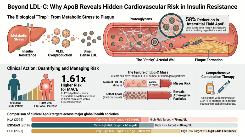
L'LDL-C misura la massa totale di colesterolo contenuta nelle particelle di LDL, piuttosto che la concentrazione delle particelle stesse.[3] Tuttavia, il contenuto di colesterolo per singola particella è altamente variabile, in quanto influenzato dalle condizioni metaboliche sistemiche e dal rimodellamento lipidico.[4] Nei pazienti con disfunzione metabolica, alti trigliceridi, oppure insulina A causa della resistenza, le particelle di LDL subiscono spesso un'esaurimento del loro nucleo di colesterolo, trasformandosi in particelle più piccole e più dense.[9] Di conseguenza, un individuo può presentare una massa di colesterolo LDL normale o addirittura bassa, pur avendo un numero molto elevato di particelle LDL piccole e dense.[9]
In tali scenari discordanti, il colesterolo LDL (LDL-C) sottostima sistematicamente il reale carico aterogenico, mentre l’ApoB quantifica correttamente il numero assoluto di particelle.[1]
Ciò non significa che l’ApoB sia universalmente superiore in ogni contesto. Il colesterolo non-HDL è un’alternativa economica, convalidata e ampiamente raccomandata dalle linee guida, che rileva tutte le lipoproteine contenenti ApoB senza richiedere un test aggiuntivo; inoltre, diverse analisi hanno evidenziato che l’ApoB, il colesterolo non-HDL e numero di particelle LDL mostrano risultati simili quando i livelli lipidici sono concordanti; il valore aggiunto dell’ApoB è massimo proprio nel fenotipo discordante e insulino-resistente che costituisce l’oggetto della presente revisione. Il consenso degli esperti propende sempre più a favore dell’ApoB come misura singola più diretta del numero di particelle aterogene, ma permane un ragionevole dibattito sul suo valore aggiunto in popolazioni non selezionate e concordanti.[13] [22]
Prove epidemiologiche causali e prospettiche
Studi genetici e osservazionali hanno chiaramente dimostrato che l’ApoB è un fattore causale di aterosclerosi piuttosto che un mero indicatore di rischio.[3]
Randomizzazione mendeliana (MR) Un europeo su larga scala studio di associazione intergenomica (GWAS) utilizzando la MR a due campioni ha dimostrato che elevazioni geneticamente determinate di ApoB sono causalmente associate a malattia coronarica (CHD) (OR 1,71, IC al 95% 1,53–1,91; P = 0,010), aterosclerosi delle grandi arterie ictus (ISL) (OR 1,43, IC 95% 1,23–1,66; P = 2,7×10⁻⁶) e ictus da piccoli vasi (ISS) (OR 1,22, IC 95% 1,06–1,41; P = 0,005).[1] La MR multivariata indica inoltre che il beneficio clinico di ipolipemizzante è fondamentalmente proporzionale alla riduzione assoluta ottenuta nel numero di particelle contenenti ApoB, piuttosto che alla massa di LDL-C rimossa.[15] [16]
Coorti prospettiche: Nelle analisi prospettiche che includono UK Biobank e nelle popolazioni in prevenzione secondaria, l'ApoB è emerso come il parametro lipidico più costantemente associato a infarto miocardico (MI) dopo la correzione per concentrazione, tipo e contenuto delle particelle.[2] Nelle popolazioni in prevenzione primaria (N = 389.529; mediana 11,1 anni), ogni aumento di 1 DS della concentrazione di ApoB è stato associato a un valore aggiustato hazard ratio pari a 1,27 (IC 95% 1,15–1,40; P < 0,001) per l’infarto miocardico incidentale, e l’ApoB è stato l’unico parametro lipidico a rimanere significativo dopo l’aggiustamento completo.[2]
Discordanza analisi Quando i livelli di ApoB e di LDL-C o non-HDL-C sono discordanti, le analisi prospettiche dimostrano che l'ApoB mantiene il valore predittivo mentre le misure del colesterolo si attenuano.[3] Nella UK Biobank (N = 41.099; 9.663 eventi MACE e 1.754 casi incidenti di CAD in circa 10 anni), l’ApoB ha dato risultati migliori rispetto al numero di particelle di LDL: in caso di discordanza 30%, l’hazard ratio ha raggiunto 1,4 per gli eventi MACE e 2,5 per la CAD, mentre un numero di particelle di LDL discordantemente elevato non ha predetto il rischio in modo indipendente.[3]
Tabella 1. Evidenze genetiche e prospettive per l'ApoB come causa cardiovascolare fattore di rischio.
| Misura di esito | Tipo di studio e fonte | Stima statistica | Significato |
| Malattia coronarica | GWAS europeo / Mendeliano randomizzazione [1] | OR 1,71 (1,53–1,91) | P = 0,010 |
| Ictus ischemico da aterosclerosi delle grandi arterie | GWAS europeo / Randomizzazione mendeliana [1] | OR 1,43 (1,23–1,66) | P = 2.7×10⁻⁶ |
| Ictus dei piccoli vasi | GWAS europeo / Randomizzazione mendeliana [1] | OR 1,22 (1,06–1,41) | P = 0,005 |
| Infarto miocardico | Coorte di prevenzione primaria [2] | aHR 1,27 (1,15–1,40) per 1-SD di ApoB | P < 0,001 |
| Rischio discorde (ApoB vs. numero di particelle LDL) | analisi di discordanza della UK Biobank [3] | HR fino a 2,5 (CAD) per la discordanza ApoB a 30% | P < 0,0001 |
2. Insulino-resistenza e prediabete: fisiopatologia della disregolazione glicemica e del dismetabolismo lipidico
L'insulino-resistenza rappresenta uno stato di ridotta risposta cellulare fisiologica all'insulina, in particolare nel tessuto muscolare scheletrico, nel tessuto adiposo e nel fegato.[4] Per superare questa resistenza, le cellule beta pancreatiche aumentano la secrezione di insulina, producendo iperinsulinemia compensatoria cronica. Nel corso del tempo, la capacità pancreatica viene sopraffatta, culminando in una progressiva disglicemia a partire da livelli normali glucosio tolleranza a prediabete e tipo 2 conclamato diabete (T2DM).[10]
La progressione della disregolazione glicemica
I dati clinici trasversali confermano che un'elevata ApoB è indipendentemente associata al peggioramento dei parametri glicemici anche in individui non diabetici.[5] Livelli più elevati di ApoB sono associati a un aumento della glicemia a digiuno (β ≈ 2,07 mg/dL per ogni deviazione standard dell’ApoB), a livelli più elevati di emoglobina glicata (β ≈ 0,06%) e a un aumento di HOMA-IR (β ≈ 0,54; tutti i P < 0,001).[5] Gli individui con i livelli più alti di ApoB quartile presentano un rischio significativamente più elevato di prediabete rispetto a coloro che si trovano nel quartile più basso (OR aggiustato 1,53, IC 95% 1,22–1,91; P < 0,001).[5]
In coorti apparentemente sane e normoglicemiche (N = 7.427), esistono correlazioni positive tra sindrome metabolica, insulino-resistenza e marker aterogeni (ApoB, colesterolo totale/HDL-C e i rapporti LDL-C/HDL-C), mentre correlazioni negative si riscontrano per gli indici cardioprotettivi come ApoA-I, ApoA-I/ApoB e HDL-C/ApoA-I.[6] Ciò sottolinea che il rimodellamento lipidico dell'insulino-resistenza è attivo e prevalente prima di qualsiasi diagnosi formale di diabete.[6]
Alterazioni del metabolismo delle lipoproteine
L'insulino-resistenza rimodella il metabolismo lipidico sistemico attraverso diversi percorsi:
- Lipolisi incontrollata: Nel tessuto adiposo insulino-resistente, l'insulina non riesce a sopprimere lipasi sensibile agli ormoni, producendo un flusso incontrollato di acidi grassi liberi (FFAs) nella circolazione portale.[7]
- Sovrapproduzione epatica di VLDL: L'afflusso di FFA al fegato stimola la sintesi e la secrezione di particelle VLDL₁ grandi e ricche di trigliceridi.[7]
- Ipertrigliceridemia: Eccessiva secrezione epatica di VLDL combinata con la downregulation della stimolazione insulinica lipoproteina lipasi (LPL) compromette la clearance di lipoproteine ricche di trigliceridi (TRL), producendo ipertrigliceridemia persistente.[7]
- Rimodellamento verso LDL piccole e dense: In condizioni di ipertrigliceridemia, estere del colesterolo trasferimento proteina (CETP) trasferisce i trigliceridi da VLDL a LDL e HDL in cambio di esteri del colesterolo. La successiva idrolisi da parte della lipasi epatica di queste particelle arricchite di trigliceridi produce LDL piccole e dense (sdLDL) e HDL instabili che vengono rapidamente depurate dal rene, deprimendo l'HDL-C.[9]
- Amplificazione delle particelle di ApoB: Poiché la secrezione epatica di VLDL aumenta e la clearance dei residui è compromessa, il numero assoluto di VLDL circolanti particelle contenenti ApoB aumenta notevolmente, anche se la massa di LDL-C rimane statica o diminuisce.[7]
Meccanismi patofisiologici sistemici
L'insulino-resistenza accelera la patologia vascolare attraverso diversi percorsi non lipidici:
- Disfunzione endoteliale: Segnalazione compromessa del recettore dell'insulina in cellule endoteliali regola al ribasso sintasi endoteliale dell'ossido nitrico (eNOS), riducendo disponibilità biologica di ossido nitrico, compromettendo la vasodilatazione e promuovendo un fenotipo endoteliale pro-coagulante e adesivo.[8]
- Stress ossidativo e cronico infiammazione: Iperglicemia e l'eccesso di lipidi guidano la sovrapproduzione mitocondriale di specie reattive dell'ossigeno (ROS), attivando fattori di trascrizione pro-infiammatori ed elevando i marker sistemici come la sensibilità elevata Proteina C-reattiva e (hsCRP) interleuchina-6 (IL-6).[10]
- Placca instabilità L'infiammazione vascolare cronica promuove macrofago infiltrazione e formazione di cellule schiumose; queste cellule secernono metalloproteinasi della matrice (MMP) che degradano il collagene cappuccio fibroso, predisponente lesioni rompere e trombosi.[8]
3. Perché livelli elevati di ApoB sono particolarmente pericolosi nei soggetti con insulino-resistenza
La coesistenza di insulino-resistenza e livelli elevati di ApoB crea un ambiente vascolare altamente distruttivo. L'insulino-resistenza non si limita ad aumentare la concentrazione di particelle aterogene di ApoB, ma altera sia le proprietà fisiche delle particelle sia la biologia strutturale della parete arteriosa, moltiplicando il rischio di intrappolamento delle particelle e il conseguente sviluppo di placca.[7]
Rimodellamento dei proteoglicani arteriosi
Negli stati di insulino-resistenza, il subendoteliale matrice extracellulare della parete arteriosa subisce un profondo rimodellamento. Insulina cronicamente elevata e citochine infiammatorie stimolano le cellule della muscolatura liscia vascolare a sovrapprodurre specifici proteoglicani, in particolare biglicano e proteoglicani di solfato di condroitina.[7] L'insulino-resistenza altera anche il processamento enzimatico di questi proteoglicani, producendo molecole più lunghe glicosaminoglicano Catene di (GAG) e aumentata solfatazione dei GAG.[7]
Poiché i GAG sono fortemente carichi negativamente, questo rimodellamento strutturale aumenta drasticamente la “viscosità” del spazio sottendoteliale, creando una trappola fisica che cattura le lipoproteine aterogeniche circolanti — una diretta estensione del modello risposta-ritenzione di aterogenesi.[7] [17]
Affinità di legame migliorata delle lipoproteine piccole e dense
Simultaneamente, le proprietà fisiche delle lipoproteine contenenti ApoB vengono modificate. Le particelle di sdLDL e i residui di VLDL predominanti nell'insulino-resistenza espongono segmenti di amminoacidi carichi positivamente sul guscio proteico di ApoB-100.[18] Questi domini positivi formano forti complessi elettrostatici con i gruppi solfato carichi negativamente sulle catene di GAG allungate dei proteoglicani arteriosi.[7]
Di conseguenza, le particelle di sdLDL e le particelle remnants mostrano un'affinità di legame significativamente maggiore per la parete vascolare rispetto alle particelle di LDL più grandi e a bassa densità.[7]
Evidenza Interstiziale di Intrappolamento Transvascolare
Per testare questo modello di intrappolamento negli esseri umani, i ricercatori hanno misurato le concentrazioni di lipoproteine nel sangue periferico liquido interstiziale rispetto al siero.[19] Poiché il liquido interstiziale drena la parete vascolare, le particelle che attraversano la endotelio senza essere intrappolato dovrebbe comparire nell'IF.[19]
Gli studi dimostrano che il rapporto tra ApoB nel liquido interstiziale e nel siero è inferiore di circa 58% nei pazienti affetti da diabete di tipo 2 rispetto ai soggetti sani di controllo (≈0,14 vs. ≈0,33).[19] Questa discrepanza fornisce prove fisiche dirette di una maggiore ritenzione transvascolare e intrappolamento delle lipoproteine contenenti ApoB all'interno della matrice subendoteliale degli individui insulino-resistenti.[19] Ulteriori studi su biopsie cutanee rivelano un livello più elevato di colesterolo non esterificato nei soggetti diabetici rispetto ai controlli, confermando l'accumulo tissutale di colesterolo.[19]
Il prolungato intrappolamento subendoteliale amplifica la cascata aterogenica: le particelle intrappolate sono esposte a enzimi ossidativi locali, sfingomielinasi segretoria (SMase) e proteasi della matrice che li modificano in LDL ossidata, inducendo il reclutamento di macrofagi e la formazione di cellule schiumose e guidando un'aterosclerosi rapida e silenziosa.[19] Questo spiega perché avanzati, placche vulnerabili può accumularsi rapidamente nei pazienti insulino-resistenti ancor prima che progrediscano verso il diabete clinico.[7]
Interazione sinergica e rischio cardiovascolare residuo
Quando l'insulino-resistenza e l'ApoB elevata si manifestano insieme, le prove meccanicistiche e precliniche suggeriscono che il loro impatto combinato sull'aterogenesi potrebbe essere amplificato anziché semplicemente additivo.[7] In condizioni metaboliche normali, un'alta concentrazione di particelle contenenti ApoB può circolare con una minore probabilità di ritenzione arteriosa. In presenza di insulino-resistenza, tuttavia, la matrice di proteoglicani alterata e la prevalenza di sdLDL altamente adesive fanno sì che anche concentrazioni moderate di ApoB circolante vengano rapidamente catturate, guidando un'aterosclerosi silente.[7]
La discordanza tra LDL-C normale e ApoB elevata
Questa interazione è più problematica quando i pazienti presentano una massa di LDL-C normale o ottimale ma livelli elevati di ApoB e insulino-resistenza sottostante.[9] Un pannello lipidico standard suggerisce un rischio basso, mentre l'aumento della conta delle particelle di ApoB combinato con la disfunzione metabolica guida il danno vascolare progressivo.[1] Il rapporto ApoB/LDL-C funge da affidabile surrogato di questo fenotipo di LDL piccole e dense ed è elevato nel diabete di tipo 2 (≈0,81 ± 0,18 vs. ≈0,74 ± 0,15 nei non diabetici; P < 0,001).[9]
Questa sinergia contribuisce notevolmente a “rischio cardiovascolare residuo”— il tasso di eventi persistenti in pazienti con diabete, sindrome metabolica o insulino-resistenza che hanno raggiunto il livello target di LDL-C con la terapia standard statina terapia.[11] Le statine sottoregolano Recettori delle LDL e eliminano le grandi particelle di LDL ricche di colesterolo, ma spesso lasciano in circolazione un'alta concentrazione di LDL piccole e dense, residui di VLDL e Lp(a).[20]
Rischio residuo è, tuttavia, multifattoriale, e contenente ApoB carico di particelle è solo uno dei suoi fattori scatenanti. La lipoproteina(a), l'infiammazione sistemica (riflessa da livelli elevati di hsCRP), ipertensione, malattia renale cronica, l'adiposità viscerale e i fattori legati allo stile di vita contribuiscono tutti indipendentemente e spesso coesistono con il fenotipo lipoproteico insulino-resistente.[11] Una strategia completa di riduzione del rischio affronta pertanto questi fattori di contribuzione insieme alla riduzione dell'ApoB, anziché trattare il carico di particelle in modo isolato.[26]
Formulazione dell'ApoB Ponderata per il Rischio
Per ovviare a questa limitazione, i ricercatori hanno formulato la metrica dell“”ApoB ponderata per il rischio" (RW-apoB), che pondera le sottrazioni contenenti ApoB (i residui di lipoproteine ricche di trigliceridi, la Lp(a) e l'ApoB delle LDL) in base alla loro aterogenicità relativa per cogliere il maggiore rischio associato alle particelle residue e modificate in un unico valore.[20]
In una coorte derivata dalla UK Biobank non sottoposta a terapia ipolipemizzante (N = 285.060), l’RW-apoB ha riclassificato circa un terzo degli individui nel suo quintile superiore rispetto alla sola ApoB misurata, identificando pazienti erroneamente classificati come a basso rischio che tuttavia presentavano un elevato tasso di eventi di CHD pari a circa 5,41 TP9T (rispetto a 3,91 TP9T nel quintile superiore dell’ApoB misurata).[20] RW-apoB ha superato costantemente l'ApoB nel predire la CHD nelle coorti trattate con statine e ad alto rischio (C-index di Harrell più elevato; P < .0001), offrendo uno strumento pratico per identificare il rischio residuo.[20]
Tabella 2. ApoB rispetto ad ApoB ponderata per il rischio (RW-apoB) per la stratificazione del rischio residuo.
| Metrica di stratificazione | Scopo clinico | Riclassificazione | tasso di eventi CHD (quintile superiore) |
| Concentrazione di ApoB [20] | Conta tutte le particelle aterogeniche | Norma di riferimento | ~3,91 TP9T (trascura i residui ad alto rischio) |
| ApoB ponderata per il rischio (RW-apoB) [20] | Pondera ApoB, residui e Lp(a) in base all'aterogenicità | Riclassificato circa 1/3 dei soggetti del quintile superiore | ~5,41 TP9T (riflette un elevato rischio residuo) |
4. Analisi quantitativa dei principali eventi cardiovascolari avversi (MACE)
Ampi studi di coorte e studi clinici hanno quantificato il valore predittivo individuale e congiunto di ApoB e dei marker di insulino-resistenza per eventi avversi cardiovascolari maggiori (MACE), infarto miocardio,ictus e mortalità.[1]
Prove da ampie coorti prospettiche
In una coorte prospettica di 11.918 partecipanti alla UK Biobank con diabete di tipo 2 e senza ASCVD al baseline, seguiti per una mediana di 185 mesi (~15,4 anni), sia l'ApoB che l“”ApoB in eccesso" (ApoB osservata meno l'ApoB predetta dall'LDL-C in una popolazione di riferimento naive alle statine) sono state associate a eventi incidenti di ASCVD e MACE — in modo lineare per l'ApoB e con una relazione a forma di J per l'ApoB in eccesso.[21]
Rispetto al valore di riferimento del 50° percentile più basso, i partecipanti con livelli più elevati di ApoB e di ApoB in eccesso hanno mostrato un marcato aumento del rischio (per ogni 1 deviazione standard di ApoB: HR 1,61, IC 95% 1,40–1,85; per ogni 1 deviazione standard di ApoB in eccesso: HR 1,18, IC 95% 1,13–1,23), in particolare tra i pazienti in terapia con statine e quelli con bassi livelli di LDL-C.[21]
In una coorte ad alto rischio di malattia coronarica I pazienti affetti da cardiopatia coronarica (CAD) seguiti per circa un decennio, il rapporto basale ApoB/LDL-C (che riflette le piccole dimensioni delle particelle LDL e la disfunzione metabolica) hanno predetto in modo significativo futuri eventi cardiovascolari indipendentemente dallo stato di diabete di tipo 2.[9]
In un modello di Cox multivariabile corretto, sia il rapporto ApoB/LDL-C che il T2DM sono risultati forti, predittori indipendenti degli eventi, con rapporti di rischio aggiustati standardizzati pari a 1,17 (IC 95% 1,05–1,30; P = 0,005) e 1,49 (IC 95% 1,26–1,75; P < 0,001), rispettivamente.[12]
Potere predittivo dei marcatori glicemici e lipoproteici
Marker clinici alternativi della resistenza all'insulina — l'indice di massa corporea trigliceridi-glucosio (TyG-BMI), l'indice trigliceridi-glucosio (TyG) e il rapporto TG/HDL-C — mostrano un forte valore prognostico per gli eventi cardiovascolari maggiori (MACE).[10] In una coorte prospettica di 1.688 pazienti con infarto miocardico prematuro (PMI) seguiti per una mediana di 17,4 mesi, i soggetti appartenenti al quartile più alto del TyG-BMI presentavano un rischio di MACE quasi triplo rispetto a quelli del quartile più basso (HR 2,88, IC 95% 1,83–4,53).[10] L’associazione risultava amplificata nei pazienti con diabete concomitante (HR 3,85, IC 95% 1,79–8,27) e in quelli con infiammazione sistemica (hsCRP elevata) (HR 3,38; IC 95% 1,78–6,43); anche l’indice TyG si è rivelato predittivo (HR 1,77, IC 95% 1,11–2,82), mentre il rapporto TG/HDL-C non ha raggiunto la significatività (HR 1,44, IC 95% 0,93–2,22).[10]
La relazione tra bassi livelli di LDL-C/ApoB e mortalità per tutte le cause
Un'osservazione epidemiologica frequentemente citata è l'associazione a U o inversa tra livelli molto bassi di colesterolo LDL e mortalità, riportata in alcune coorti della popolazione generale e di anziani, in cui un basso livello di LDL-C si associa a fragilità, malnutrizione e malattie croniche.[15] In un'analisi del NHANES statunitense (2005–2016; N = 15.380; follow-up mediano di 101 mesi; 1.771 decessi), sia la bassa ApoB che il basso LDL-C sono stati associati a più alto mortalità per tutte le cause e mortalità cardiovascolare: rispetto a un livello di ApoB < 90 mg/dL, un livello di ApoB ≥ 90 mg/dL presentava un rapporto di rischio (hazard ratio) pari a 0,79 (IC 95% 0,69–0,89) per mortalità per tutte le cause.[15]
Cautela interpretativa. Questo segnale inverso è ampiamente attribuito a causalità inversa e confondente da fragilità e malattia subclinica in popolazioni non selezionate, piuttosto che da un effetto protettivo di un elevato carico di particelle aterogene. Ciò non contraddice la relazione causale e dose-dipendente tra ApoB e aterosclerosi stabilita dalla randomizzazione mendeliana e dagli studi clinici randomizzati di riduzione dei lipidi.[1] [16] Nelle popolazioni trattate con statine e in prevenzione secondaria, l'ApoB rimane un indicatore diretto e positivo del rischio cardiovascolare residuo e di mortalità, motivo per cui la riduzione dell'ApoB — e non semplicemente della massa di LDL-C — costituisce l'obiettivo terapeutico.[22]
Tabella 3. Stime del rischio per l'ApoB e i marker di insulino-resistenza nelle coorti cliniche.
| Coorte clinica | Analizzato biomarcatori | Stima del rischio corretta (intervallo di confidenza 95%) | Endpoint |
| UK Biobank, T2DM [21] | ApoB vs. ApoB in eccesso (per 1-SD) | ApoB HR 1,61 (1,40–1,85); eccesso di ApoB HR 1,18 (1,13–1,23) | Incidenti ASCVD (2.548) & MACE (1.205) |
| CAD ad alto rischio [12] | Rapporto ApoB/LDL-C e stato del diabete | ApoB/LDL-C HR 1,17 (1,05–1,30, P=0,005); T2DM HR 1,49 (1,26–1,75, P<0,001) | Eventi CV in circa 10 anni |
| Infarto miocardico precoce [10] | TyG-BMI, TyG, TG/HDL-C (Q4 vs Q1) | TyG-BMI HR 2,88 (1,83–4,53); TyG 1,77 (1,11–2,82); TG/HDL-C 1,44 (0,93–2,22, NS) | Incidente MACE in 17,4 mesi |
| NHANES degli Stati Uniti [15] | Siero ApoB / LDL-C | ApoB ≥90 vs <90 mg/dL: HR 0,79 (0,69–0,89) — ApoB basso è associato a una maggiore mortalità (paradosso causalità) | Mortalità per tutte le cause (~8,4 anni) |
5. Linee guida e stratificazione del rischio: integrazione di ApoB e insulino-resistenza
Riconoscendo il superiore valore predittivo dell'ApoB rispetto ai lipidi tradizionali, le principali società cardiologiche e lipidologiche hanno integrato la misurazione dell'ApoB e i relativi valori target nelle loro linee guida per la stratificazione del rischio, in particolare per i pazienti con disfunzione metabolica.[11]
Confronto tra linee guida di consenso e obiettivi
Le società scientifiche offrono raccomandazioni diverse in base al profilo di rischio del paziente:
Associazione Nazionale dei Lipidi (NLA): Il consenso degli esperti NLA del 2024 sull'ApoB raccomanda soglie di trattamento di circa 60 mg/dL (rischio molto alto), circa 70 mg/dL (rischio alto) e circa 90 mg/dL (rischio borderline-intermediario), e classifica l'ApoB ≥130 mg/dL (circa 90° percentile) come fattore che aumenta il rischio. L'ApoB è raccomandata per la valutazione iniziale del rischio cardiovascolare o come obiettivo secondario opzionale.[23]
Società Europea di Cardiologia / Società Europea dell'Aterosclerosi (ESC/EAS): Le linee guida ESC/EAS del 2019 fissano obiettivi secondari di ApoB <65 mg/dL (rischio molto alto), <80 mg/dL (rischio alto) e <100 mg/dL (rischio moderato), corrispondenti a obiettivi di LDL-C <55, <70 e <100 mg/dL. Esse approvano l'ApoB come misurazione di screening primario alternativa, in particolare nei pazienti con sindrome metabolica, diabete di tipo 2, obesità, ipertrigliceridemia o LDL-C molto basso.[24]
Società Cardiovascolare Canadese (CCS): Le linee guida CCS del 2021 preferiscono ApoB o non-HDL-C rispetto a LDL-C quando i trigliceridi superano 1,5 mmol/L (~133 mg/dL), sia nella prevenzione primaria che prevenzione secondaria, con soglie di intensificazione di ApoB ≥0,8 g/L (aggiungere ezetimibe) e ≥0,7 g/L (ulteriore intensificazione).[25]
American College of Cardiology / American Heart Association (ACC/AHA): Il Linee guida multisocietarie ACC/AHA sulla dislipidemia del 2026 adotta le equazioni PREVENT-ASCVD per stimare il rischio a 10 e 30 anni negli adulti di età compresa tra i 30 e i 79 anni senza ASCVD. Gli obiettivi per il colesterolo LDL (LDL-C) sono <100 mg/dL (rischio borderline/intermedio), <70 mg/dL (rischio elevato) e <55 mg/dL (prevenzione secondaria per rischio molto elevato). La misurazione dell’ApoB è ragionevole (Classe 2a) negli adulti in terapia ipolipemizzante — in particolare quelli con ASCVD, sindrome cardiovascolare-renale-metabolica (CKM), T2DM o trigliceridi elevati — per orientare l’intensificazione della terapia una volta raggiunti gli obiettivi di LDL-C e non-HDL-C.[26]
Tabella 4. Confronto tra i target di ApoB delle società scientifiche e applicazione raccomandata.
| Società | Rischio altissimo / prevenzione secondaria | Alto rischio / prevenzione primaria | Applicazione consigliata |
| NLA [23] | ApoB ~60 mg/dL | ApoB ~70 mg/dL (90 mg/dL borderline-intermedio) | Obiettivo secondario opzionale; ≥130 mg/dL è un fattore che aumenta il rischio. |
| ESC/EAS [24] | ApoB <65 mg/dL | ApoB <80 mg/dL (<100 moderato) | Bersaglio diagnostico/di screening primario alternativo; preferito nella sindrome metabolica, nel diabete mellito di tipo 2 (T2DM), nell'obesità. |
| CCS [25] | ApoB ≥0,7 g/L (intensificare) | ApoB ≥0,8 g/L (aggiungere ezetimibe) | Preferito rispetto al C-LDL quando i TG >1,5 mmol/L (~133 mg/dL). |
| ACC/AHA [26] | In linea con LDL-C <55 mg/dL | LDL-C <70 mg/dL (alto); <100 mg/dL (intermedio) | Classe 2a; guide all'intensificazione dopo il raggiungimento degli obiettivi di LDL-C nella sindrome CKM, nel diabete e con alti livelli di TG. |
Criteri di misurazione di routine nelle popolazioni ad alto rischio
Esiste una forte logica clinica per misurare routinariamente l'ApoB nei pazienti con insulino-resistenza, obesità, prediabete o sindrome metabolica.[11] In queste popolazioni, l'iperproduzione epatica di VLDL e il rimodellamento lipidico producono un'alta concentrazione di particelle LDL piccole e dense.[7]
Poiché queste particelle trasportano meno colesterolo per particella, le misurazioni standard di LDL-C possono sembrare normali o basse, mascherando un'alta concentrazione di particelle aterogene.[1] La misurazione diretta dell'ApoB quantifica questo carico di particelle, identificando gli individui ad alto rischio che altrimenti verrebbero classificati erroneamente come a basso rischio dai tradizionali pannelli lipidici.[1]
Questa utilità è riconosciuta nelle linee guida sulle dislipidemie ACC/AHA, che incorporano le equazioni PREVENT-ASCVD per stimare il rischio cardiovascolare a 10 e 30 anni e raccomandano la misurazione dell'ApoB (Classe 2a) per valutare il rischio residuo e guidare l'intensificazione nei pazienti con sindrome CKM, diabete di tipo 2 o trigliceridi elevati che hanno già raggiunto i loro obiettivi di LDL-C e non-HDL-C.[26]
6. Implicazioni Cliniche e Percorsi Terapeutici
In un paziente insulino-resistente, un'ApoB elevata può indicare un rischio aterogeno residuo maggiore rispetto a quello espresso dal solo LDL-C; tuttavia, l'intensità del trattamento dovrebbe essere guidata dal rischio ASCVD complessivo e dalle indicazioni definite dalle linee guida, piuttosto che presupporre l'equivalenza con il diabete di tipo 2 conclamato.[11] La fisiopatologia sottostante — il rimodellamento vascolare-matrice, la disfunzione endoteliale e l'accelerata ritenzione subendoteliale delle particelle contenenti ApoB — è meccanisticamente attivo durante le fasi insulino-resistente e prediabetica ed è supportato da studi meccanicistici e preclinici, il che può guidare aterosclerosi subclinica prima che il diabete venga diagnosticato clinicamente.[7]
Statine e la sfida del rischio residuo
Le statine inibiscono la sintesi epatica Idrossi-metilglutaril-CoA reduttasi, riducendo la concentrazione intracellulare sintesi del colesterolo e sovraregolando i recettori epatici delle LDL, il che migliora la clearance delle particelle circolanti contenenti ApoB.[27] Statine ad alta intensità sono raccomandati per ottenere una sostanziale riduzione del C-LDL.[26] Tuttavia, nei pazienti insulino-resistenti, le statine possono lasciare un'elevata concentrazione di particelle residue piccole e dense di LDL e di residui, sottolineando la necessità di una terapia di combinazione.[11]
Terapie ipolipemizzanti non statiniche mirate
Le terapie non statiniche mirate riducono ulteriormente l'ApoB e diminuiscono gli eventi cardiovascolari:
- Ezetimibe: Inibendo NPC1L1 trasportatore nell'intestino tenue, l'ezetimibe riduce l'assorbimento del colesterolo.[27] Nei modelli animali insulino-resistenti, l'ezetimibe — da solo o con simvastatina — ha ridotto la ritenzione arteriosa ex vivo di lipoproteine remnant di ApoB-48 e ApoB-100 di origine intestinale, limitando il deposito di colesterolo.[28]
- Inibitori di PCSK9: Questa classe comprende gli anticorpi monoclonali alirocumab e evolocumab (e il nuovo agente tafolecimab) e il piccolo RNA interferente inclisiran.[29] Impedendo la degradazione dei recettori delle LDL mediata dalla PCSK9, questi agenti preservano il riciclo dei recettori, riducendo il colesterolo LDL (LDL-C) di circa il 50–60%, con corrispondenti riduzioni dell’ApoB.[29] L'orale PCSK9 l'inibitore enlicitide decanoato (20 mg una volta al giorno) ha ridotto il colesterolo LDL di circa 55,8% rispetto a placebo alla 24ª settimana dello studio di fase 3 CORALreef Lipids, con una concomitante riduzione di ApoB.[30]
- ANGPTL3 inibitori La proteina simile all'angiopoietina 3 (ANGPTL3) regola il metabolismo dei lipidi plasmatici inibendo la lipoproteina lipasi e la lipasi endoteliale.[31] Evinacumab, un anticorpo monoclonale contro l’ANGPTL3 (un bersaglio diverso dal PCSK9), riduce il colesterolo LDL di circa il 47–49% nei pazienti con ipercolesterolemia refrattaria ipercolesterolemia familiare omozigote (HoFH).[31] Le terapie emergenti a base di oligonucleotidi antisenso (vupanorsen) e siRNA (zodasiran, solbinsiran) agiscono sul metabolismo dei trigliceridi, ottenendo riduzioni dei trigliceridi superiori a 50%, accompagnate da modeste riduzioni del colesterolo LDL (≤16%).[32] Nello studio TRANSLATE-TIMI 70, il vupanorsen non ha prodotto alcun beneficio chiaro, dose-dipendente, in termini di LDL-C/ApoB ed è stato associato ad aumenti dose-dipendenti del contenuto di grasso epatico (aumento relativo fino a ~76%), determinando l'interruzione del suo sviluppo.[32]
- Acido bempedoico: Un esame orale profamaco attivato specificamente nel fegato da ACSVL1 — un enzima assente nel muscolo scheletrico, evitando così la miopatia associata alle statine — che inibisce ATP-citrato liasi (ACLY) a monte della HMG-CoA reduttasi.[27]
Nel Risultati CLEAR studio clinico (13.970 pazienti intolleranti alle statine affetti da ASCVD o ad alto rischio di ASCVD), l'acido bempedoico (180 mg una volta al giorno) ha ridotto il colesterolo LDL di circa 21,1% e la hsCRP di circa 22,2% a 6 mesi.[33] Per una mediana di 40,6 mesi, ha ridotto l'endpoint primario MACE a 4 componenti (morte CV, infarto miocardico non fatale, ictus non fatale o coronarica rivascolarizzazione) di 13% (HR 0,87, IC 95% 0,79–0,96; P = 0,004).[33] Ha inoltre ridotto gli eventi cardiovascolari maggiori (MACE) a tre componenti (HR 0,85, 0,76–0,96), l’infarto miocardico (HR 0,77; 0,66–0,91) e la rivascolarizzazione coronarica (HR 0,81; 0,72–0,92); nel sottogruppo di prevenzione primaria l’HR per gli eventi MACE a 4 componenti era pari a 0,70 (IC 95% 0,55–0,89).[33]
Modifica metabolica basata sulle incretine
Le terapie a base di incretina come semaglutide e tirzepatide ottenere perdita di peso e migliorare i profili di rischio cardiovascolare.[34]
- Semaglutide Nel SELEZIONA prova (N = 17.604 pazienti in sovrappeso o obesi con malattie cardiovascolari preesistenti ma senza diabete), il semaglutide alla dose di 2,4 mg una volta alla settimana ha ridotto gli eventi cardiovascolari maggiori (MACE) a 3 punti del 20% (HR 0,80, 95% IC 0,72–0,90; P < 0,001) nell’arco di una durata media di 39,8 mesi. Nelle analisi dello studio SELECT sono state riportate riduzioni dei marcatori infiammatori e aterogenici (tra cui hsCRP e ApoB), ma tali risultati erano secondari al beneficio primario in termini di esiti cardiovascolari e dovrebbero essere riferiti a quelle specifiche analisi.[34]
- Tirzepatide: Nel SURMOUNT-1 Nel corso dello studio, il tirzepatide ha determinato una perdita di peso fino a circa 20,91 TP9T e ha migliorato sensibilità all'insulina.[35] In uno studio di fase 2b sull’analisi delle lipoproteine nel diabete di tipo 2, il tirzepatide ha ridotto le particelle di lipoproteine di grandi dimensioni ricche di trigliceridi, le particelle di LDL di piccole dimensioni e il punteggio di insulino-resistenza delle lipoproteine (LPIR), con riduzioni dell’ApoB fino a ~17% e riduzioni dei trigliceridi di ~31–35%, in linea con una correzione metabolica sistemica e a livello delle particelle.[36]
- Inibitori di SGLT2: Promuovendo la glicosuria e la diuresi osmotica, gli inibitori di SGLT2 migliorano controllo glicemico, riducono il peso corporeo e il grasso e diminuiscono la mortalità cardiovascolare e renale; inoltre spostano l'utilizzazione dei substrati da carboidrato verso l'ossidazione dei lipidi e dei corpi chetonici.[37]
- Interventi sulla dieta e sullo stile di vita: È raccomandato l'invio a un dietista nutrizionista registrato (RDN) per i pazienti con trigliceridi elevati (TG a digiuno ≥1000 mg/dL, COR 1; 150–999 mg/dL con sindrome CKM, COR 2a) al fine di fornire una consulenza basata sulle prove di efficacia e ridurre il rischio di pancreatite.[26]
Tabella 5. Riepilogo delle terapie ipolipemizzanti e metaboliche rilevanti per la riduzione dell'ApoB.
| Agente | Efficacia su ApoB / lipidi | Processo | Risultato | Sicurezza / tollerabilità |
| Acido bempedoico [33] | LDL-C −21,1%, hsCRP −22,2% | CLEAR Outcomes (13.970 pazienti con intolleranza alle statine) | MACE-4 HR 0,87 (0,79–0,96); prevenzione primaria 0,70 (0,55–0,89) | Sintomi muscolari ≈ placebo; lieve ↑ acido urico, gotta, colelitiasi |
| Semaglutide [34] | Riduzione dei MACE; gli effetti sui biomarcatori non sono l'endpoint primario | SELEZIONARE (17.604 CVD obesi, niente diabete) | MACE a 3 punti HR 0,80 (0,72–0,90) | Ben tollerato; effetti gastrointestinali comuni; perdita di peso sostenuta |
| Tirzepatide [36] | ApoB fino a ~17%↓; TG ~31–35%↓; ↓LPIR | SURMOUNT / SURPASS / fase 2b | ↓ Punteggio LPIR, piccolo LDLP, grande TRLP | Effetti collaterali gastrointestinali; perdita di peso fino a ~20,91 TP9T |
| Evinacumab (anti-ANGPTL3) [31] | LDL-C ≈47–49%↓ nell’HoFH | ELIPSE HoFH | Marcata riduzione del C-LDL nella HoFH refrattaria | Ben tollerato; somministrazione endovenosa |
| Vupanorsen (ANGPTL3 ASO) [32] | TG ≈41–57%↓; LDL-C ≤16%↓ | TRANSLATE-TIMI 70 | Nessun LDL-C/ApoB chiaro dose-risposta; sviluppo interrotto | Aumento dose-dipendente del grasso epatico (relativo fino a ~76%) |
7. Discussione e conclusione
Questa analisi dimostra che la valutazione del pericolo cardiovascolare richiede di guardare oltre la semplice massa della concentrazione lipidica.[4] Quando la resistenza all'insulina e livelli elevati di ApoB coesistono, essi guidano un'aterogenesi silenziosa e progressiva attraverso diversi pathway metabolici e vascolari interconnessi.[7]
Figura 1. Percorso integrato dallo stress metabolico cronico agli eventi cardiovascolari acuti.
| STRESS METABOLICO CRONICO (Insulino-resistenza) |
▼
| Via di disregolazione lipidica
(Eccesso di VLDL epatiche) |
Percorso del rimodellamento vascolare
(Danno endoteliale) |
▼
| Conteggio elevato di ApoB
(sdLDL e residui) |
Proteoglicano intimale
Rimodellamento (GAG appiccicosi) |
▼
| RITENZIONE TRANSVASCOLARE ACCELERATA E INTRAPPOLAMENTO SUBENDOTELIALE
(rapporto IF/ApoB sierica inferiore di circa 58% nel diabete di tipo 2) |
▼
| MODIFICAZIONE OSSIDATIVA E RECLUTAMENTO DEI MONOCITI |
▼
| FORMAZIONE DI CELLULE SCHIUMOSE E PROGRESSIONE DELLA PLACCA |
▼
| rottura della placca E TROMBOSI |
▼
| EVENTI CARDIOVASCOLARI ACUTI (MACE, Infarto Miocardico, ictus) |
Discussione: un modello unificato e i suoi limiti
Le prove qui esaminate convergono su un unico modello coerente, riassunto nella Figura 1. La resistenza all'insulina cronica guida la sovrapproduzione epatica di VLDL ricche di trigliceridi e il loro rimodellamento in LDL piccole e dense, aumentando il numero di particelle di ApoB circolanti anche quando la massa di LDL-C appare normale; parallelamente, la parete arteriosa insulino-resistente viene rimodellata in una superficie più adesiva e ricca di proteoglicani che trattiene preferenzialmente tali particelle. La convergenza di particelle più aterogeniche e di una parete vascolare più ritentiva offre una spiegazione unificante del motivo per cui il rischio cardiovascolare negli individui insulino-resistenti e prediabetici può superare quanto previsto da un profilo lipidico standard.
Due cautelezioni interpretative derivano direttamente dalla forza delle prove sottostanti. Primo, i dati umani sono più forti per quanto riguarda associazioni — la causalità della randomizzazione mendeliana di ApoB, i rapporti di rischio prospettici e il segnale di ritenzione nel fluido interstiziale — mentre la sequenza graduale di rimodellamento dei proteoglicani e formazione di cellule schiumose si basa sostanzialmente su studi meccanicistici e preclinici, incluso un modello animale. Questi passaggi meccanicistici sono biologicamente plausibili e supportati, ma non sono dimostrati allo stesso livello negli umani e sono presentati qui come un meccanismo plausibile piuttosto che come un fatto clinico consolidato. Secondo, l'ApoB è uno dei fattori di rischio residuo tra diversi; lipoproteina(a), infiammazione, ipertensione, cronica malattia renale, l'adiposità e lo stile di vita agiscono indipendentemente e dovrebbero essere gestiti insieme ad esso.
Conclusione e traduzione clinica
Per il paziente insulino-resistente, il messaggio pratico è che l'LDL-C da solo può sottostimare il carico aterogeno, e che l'ApoB (o, laddove l'ApoB non sia disponibile, il non-HDL-C) coglie in modo più diretto il numero di particelle implicato dalla biologia. Tradurre tutto ciò nell'assistenza clinica non richiede di trattare il prediabete come se fosse un diabete conclamato; richiede di misurare la cosa giusta e di agire sul rischio complessivo.
Quattro principi riassumono la traslazione clinica:
- Misurare il carico di particolato. Prendere in considerazione l'ApoB (o il colesterolo non-HDL) nei pazienti con insulino-resistenza, obesità, prediabete o sindrome metabolica, nei quali il colesterolo LDL sottostima più spesso il numero di particelle aterogene.[11]
- Adatta l'intensità al rischio complessivo. Utilizzare l'ApoB elevato per perfezionare — anziché sostituire — la valutazione del rischio basata sulle linee guida, intensificando la riduzione dei lipidi in base al rischio ASCVD complessivo e alle indicazioni definite dalle società scientifiche, anziché ipotizzare un rischio equivalente a quello del diabete.[11] [26]
- Ridurre l'ApoB con terapia di combinazione ove indicato. Le statine ad alta intensità abbinate a ezetimibe, inibitori di PCSK9 o acido bempedoico massimizzano la clearance delle particelle contenenti ApoB e riducono gli eventi nelle popolazioni appropriate.[27] [33]
- Affrontare il substrato metabolico e i co-fattori. agonisti dei recettori del GLP-1 come il semaglutide riduce gli eventi cardiovascolari nell'obesità con patologia cardiovascolare accertata,[34] e il tirzepatide, un doppio agonista del GIP/GLP-1, migliora la sensibilità all'insulinica e i marcatori lipoproteici di insulino-resistenza;[36] questi, insieme al controllo del rischio correlato alla Lp(a), all'infiammazione, pressione sanguigna, e stile di vita, completare una strategia di riduzione del rischio che consideri il carico di particelle come una sola componente anziché il totale.
In sintesi, la tesi centrale sostiene che negli stati di insulino-resistenza, il carico di particelle definite da ApoB e la biologia della ritenzione arteriosa catturano insieme un rischio che il colesterolo LDL può mancare. La risposta appropriata è una migliore misurazione e un trattamento multifattoriale proporzionato al rischio, affermato con sicurezza laddove le prove randomizzate e genetiche lo supportano, e con la dovuta cautela laddove il meccanismo rimane inferenziale.
Riferimenti
Le citazioni seguono la numerazione IEEE. Questa lista di riferimenti dà priorità a studi primari revisionati da pareri pari, documenti di linee guida ufficiali e rapporti di studi indicizzati, e documenti di revisione o di consenso laddove un rapporto primario completo non fosse la fonte più appropriata; questi sono identificati come tali.
- Kong X, Cai Y, Li Y, Wang P. Causal relationship between apolipoprotein B and risk of atherosclerotic cardiovascular disease: a mendelian randomization analysis. Health Inf Sci Syst. 2025;13(1):13. Published 2025 Jan 3. doi:10.1007/s13755-024-00323-5
- Marston NA, Giugliano RP, Melloni GEM, et al. Association of Apolipoprotein B-Containing Lipoproteins and Risk of Myocardial Infarction in Individuals With and Without Atherosclerosis: Distinguishing Between Particle Concentration, Type, and Content. JAMA Cardiol. 2022;7(3):250-256. doi:10.1001/jamacardio.2021.5083
- Epstein E, Ekpo E, Evans D, et al. Apolipoprotein B outperforms low density lipoprotein particle number as a marker of cardiovascular risk in the UK Biobank. Eur J Prev Cardiol. Published online September 1, 2025. doi:10.1093/eurjpc/zwaf554
- Borén J, Chapman MJ, Krauss RM, et al. Low-density lipoproteins cause atherosclerotic cardiovascular disease: pathophysiological, genetic, and therapeutic insights: a consensus statement from the European Atherosclerosis Society Consensus Panel. Eur Heart J. 2020;41(24):2313-2330. doi:10.1093/eurheartj/ehz962
- Bekbossynova M, Ivanova-Razumova T, Kali A, et al. Apolipoprotein B and Glycemic Dysregulation: New Predictors of Type 2 Diabetes in High-Cardiovascular-Risk Populations. J Pers Med. 2025;15(5):163. Published 2025 Apr 23. doi:10.3390/jpm15050163
- Sung KC, Hwang ST. Association between insulin resistance and apolipoprotein B in normoglycemic Koreans. Atherosclerosis. 2005;180(1):161-169. doi:10.1016/j.atherosclerosis.2004.11.009
- Mangat R, Warnakula S, Borthwick F, et al. Arterial retention of remnant lipoproteins ex vivo is increased in insulin resistance because of increased arterial biglycan and production of cholesterol-rich atherogenic particles that can be improved by ezetimibe in the JCR:LA-cp rat. J Am Heart Assoc. 2012;1(5):e003434. doi:10.1161/JAHA.112.003434
- Borén J, Williams KJ. The central role of arterial retention of cholesterol-rich apolipoprotein-B-containing lipoproteins in the pathogenesis of atherosclerosis: a triumph of simplicity. Curr Opin Lipidol. 2016;27(5):473-483. doi:10.1097/MOL.0000000000000330
- BARBARA LARCHER, THOMAS PLATTNER, ARTHUR MADER, ALEXANDER VONBANK, ANDREAS LEIHERER, AXEL MUENDLEIN, HEINZ DREXEL, CHRISTOPH H. SAELY; 456-P: The ApoB/LDL-C Ratio Predicts Cardiovascular Events in Coronary Artery Disease Patients Independent of Type 2 Diabetes Status. Diabetes 14 June 2024; 73 (Supplement_1): 456–P. https://doi.org/10.2337/db24-456-P
- Zhou Y, Wang Y, Chen J, Jiang L, Chu R, Tian W, Wang J, Liu Y and Gao J (2026) The association between insulin resistance indices and the occurrence of major adverse cardiovascular events in patients with premature myocardial infarction: a prospective cohort study. Front. Nutr. 13:1724362. doi: 10.3389/fnut.2026.1724362
- Soffer DE, Marston NA, Maki KC, et al. Role of apolipoprotein B in the clinical management of cardiovascular risk in adults: An Expert Clinical Consensus from the National Lipid Association. J Clin Lipidol. 2024;18(5):e647-e663. doi:10.1016/j.jacl.2024.08.013
- BARBARA LARCHER, MAXIMILIAN MAECHLER, LUKAS SPRENGER, ARTHUR MADER, ALEXANDER VONBANK, THOMAS PLATTNER, ANDREAS LEIHERER, AXEL MUENDLEIN, HEINZ DREXEL, CHRISTOPH H. SAELY; 260-OR: The ApoB/LDL-C Ratio Predicts Major Cardiovascular Events in Cardiovascular Disease Patients Independent of Type 2 Diabetes Status. Diabetes 20 June 2023; 72 (Supplement_1): 260–OR. https://doi.org/10.2337/db23-260-OR
- Sniderman AD, Thanassoulis G, Glavinovic T, et al. Apolipoprotein B Particles and Cardiovascular Disease: A Narrative Review. JAMA Cardiol. 2019;4(12):1287-1295. doi:10.1001/jamacardio.2019.3780
- Ference BA, Ginsberg HN, Graham I, et al. Low-density lipoproteins cause atherosclerotic cardiovascular disease. 1. Evidence from genetic, epidemiologic, and clinical studies. A consensus statement from the European Atherosclerosis Society Consensus Panel. Eur Heart J. 2017;38(32):2459-2472. doi:10.1093/eurheartj/ehx144
- Yu X, Yuan Y, Dong X, et al. Low apolipoprotein B and LDL-cholesterol are associated with the risk of cardiovascular and all-cause mortality: a prospective cohort. Ann Med. 2025;57(1):2529565. doi:10.1080/07853890.2025.2529565
- Richardson TG, Sanderson E, Palmer TM, et al. Evaluating the relationship between circulating lipoprotein lipids and apolipoproteins with risk of coronary heart disease: A multivariable Mendelian randomisation analysis. PLoS Med. 2020;17(3):e1003062. Published 2020 Mar 23. doi:10.1371/journal.pmed.1003062
- Williams KJ, Tabas I. The response-to-retention hypothesis of early atherogenesis. Arterioscler Thromb Vasc Biol. 1995;15(5):551-561. doi:10.1161/01.atv.15.5.551
- Camejo G, Hurt-Camejo E, Wiklund O, Bondjers G. Association of apo B lipoproteins with arterial proteoglycans: pathological significance and molecular basis. Atherosclerosis. 1998;139(2):205-222. doi:10.1016/s0021-9150(98)00107-5
- Björklund P, Härdfeldt J, Äikäs L, et al. Increased transvascular retention of atherogenic lipoproteins in type 2 diabetes relates to their enhanced proteoglycan binding. JCI Insight. 2026;11(10):e177849. Published 2026 Apr 7. doi:10.1172/jci.insight.177849
- Rehman MB, Björnson E, Adiels M, et al. Risk-weighted apoB: a novel summary metric outperforming traditional lipid biomarkers in predicting coronary heart disease. Eur Heart J. Published online January 22, 2026. doi:10.1093/eurheartj/ehaf1124
- Lyu L, Kao C, Su J, et al. Association of apolipoprotein B and excess apolipoprotein B with cardiovascular risk in type 2 diabetes: a prospective cohort study of the UK Biobank. Lipids Health Dis. 2026;25(1):55. Published 2026 Jan 15. doi:10.1186/s12944-025-02852-8
- Sniderman AD, Navar AM, Thanassoulis G. Apolipoprotein B vs Low-Density Lipoprotein Cholesterol and Non-High-Density Lipoprotein Cholesterol as the Primary Measure of Apolipoprotein B Lipoprotein-Related Risk: The Debate Is Over. JAMA Cardiol. 2022;7(3):257-258. doi:10.1001/jamacardio.2021.5080
- Soffer DE, Marston NA, Maki KC, et al. Role of apolipoprotein B in the clinical management of cardiovascular risk in adults: An Expert Clinical Consensus from the National Lipid Association. J Clin Lipidol. 2024;18(5):e647-e663. doi:10.1016/j.jacl.2024.08.013
- Mach F, Baigent C, Catapano AL, et al. 2019 ESC/EAS Guidelines for the management of dyslipidaemias: lipid modification to reduce cardiovascular risk. Eur Heart J. 2020;41(1):111-188. doi:10.1093/eurheartj/ehz455
- Pearson GJ, Thanassoulis G, Anderson TJ, et al. 2021 Canadian Cardiovascular Society Guidelines for the Management of Dyslipidemia for the Prevention of Cardiovascular Disease in Adults. Can J Cardiol. 2021;37(8):1129-1150. doi:10.1016/j.cjca.2021.03.016
- Blumenthal RS, Morris PB, Gaudino M, et al. 2026 ACC/AHA/AACVPR/ABC/ACPM/ADA/AGS/APhA/ASPC/NLA/PCNA Guideline on the Management of Dyslipidemia: A Report of the American College of Cardiology/American Heart Association Joint Committee on Clinical Practice Guidelines. J Am Coll Cardiol. 2026;87(19):2624-2757. doi:10.1016/j.jacc.2025.11.016
- Albosta M, Grant JK, Michos ED. Bempedoic Acid: Lipid Lowering for Cardiovascular Disease Prevention. Heart Int. 2023;17(2):27-34. Published 2023 Nov 1. doi:10.17925/HI.2023.17.2.1
- Mangat R, Warnakula S, Borthwick F, et al. Arterial retention of remnant lipoproteins ex vivo is increased in insulin resistance because of increased arterial biglycan and production of cholesterol-rich atherogenic particles that can be improved by ezetimibe in the JCR:LA-cp rat. J Am Heart Assoc. 2012;1(5):e003434. doi:10.1161/JAHA.112.003434
- Sabatine MS, Giugliano RP, Keech AC, et al. Evolocumab and Clinical Outcomes in Patients with Cardiovascular Disease. N Engl J Med. 2017;376(18):1713-1722. doi:10.1056/NEJMoa1615664
- Ballantyne CM, Gellis L, Tardif JC, et al. Efficacy and Safety of Oral PCSK9 Inhibitor Enlicitide in Adults With Heterozygous Familial Hypercholesterolemia: A Randomized Clinical Trial. JAMA. 2026;335(2):129-139. doi:10.1001/jama.2025.20620
- Raal FJ, Rosenson RS, Reeskamp LF, et al. Evinacumab for Homozygous Familial Hypercholesterolemia. N Engl J Med. 2020;383(8):711-720. doi:10.1056/NEJMoa2004215
- Bergmark BA, Marston NA, Bramson CR, et al. Effect of Vupanorsen on Non-High-Density Lipoprotein Cholesterol Levels in Statin-Treated Patients With Elevated Cholesterol: TRANSLATE-TIMI 70. Circulation. 2022;145(18):1377-1386. doi:10.1161/CIRCULATIONAHA.122.059266
- Nissen SE, Lincoff AM, Brennan D, et al. Bempedoic Acid and Cardiovascular Outcomes in Statin-Intolerant Patients. N Engl J Med. 2023;388(15):1353-1364. doi:10.1056/NEJMoa2215024
- Lincoff AM, Brown-Frandsen K, Colhoun HM, et al. Semaglutide and Cardiovascular Outcomes in Obesity without Diabetes. N Engl J Med. 2023;389(24):2221-2232. doi:10.1056/NEJMoa2307563
- Jastreboff AM, Aronne LJ, Ahmad NN, et al. Tirzepatide Once Weekly for the Treatment of Obesity. N Engl J Med. 2022;387(3):205-216. doi:10.1056/NEJMoa2206038
- Wilson JM, Nikooienejad A, Robins DA, et al. The dual glucose-dependent insulinotropic peptide and glucagon-like peptide-1 receptor agonist, tirzepatide, improves lipoprotein biomarkers associated with insulin resistance and cardiovascular risk in patients with type 2 diabetes. Diabetes Obes Metab. 2020;22(12):2451-2459. doi:10.1111/dom.14174
- Szekeres Z, Toth K, Szabados E. The Effects of SGLT2 Inhibitors on Lipid Metabolism. Metabolites. 2021;11(2):87. Published 2021 Feb 1. doi:10.3390/metabo11020087

